Category Archives: MSI

Assessment in Enhanced Imaging of Cultural Heritage

In what now seems like the way distant past, just before the library building closed, Ryan Baumann and I virtually presented on DUL’s work with multispectral imaging at the More than Meets the Eye conference, hosted by the University of Iowa. The three-day conference was a wonderful opportunity to hear examples from around the world on the application of enhanced digital imaging technologies in research on cultural heritage.

This month the library kicked off a weekly “Lunch & Learn” series, in which library staff give short (~20 min) presentations about their recent work or research interests. It provides an additional opportunity to connect with our colleagues and learn something new each week.  Since I already had my slides from the Iowa conference, I volunteered to present in the first session, and today I would like to share those slides with Bitstreams readers:

Assessment in Enhanced Imaging of Cultural Heritage

I’ve included my script in the notes for each slide, so you can get the full context. There are also links to other talks the MSI team has done over the years and other work in enhanced imaging from my colleagues.

Have a safe Memorial Day weekend!

Congratulations and farewell to Mike Adamo

This week, Digitization Specialist Mike Adamo will move on from Duke Libraries after 14 years to assume a new position as Digital Imaging Coordinator at the Libraries of Virginia Tech University. Mike has contributed so much to our Digital Collections program during his tenure, providing years of uncompromising still imaging services, stewardship in times of change for the Digital Production Center, as well as leadership of and then years of service on our Digital Collections Implementation Team. He has also been the lead digitization specialist on some of our most well known digital collections like the Hugh Mangum photographs, James Karales photographs and William Gedney collection.

In addition, Mike has been a principal figure on our Multispectral Imaging Team and has been invaluable to our development of this service for the library. He established the setup and led all MSI imaging sessions; collaborated cross-departmentally with other members on the MSI Team to vet requests and develop workflows; and worked with vendors and other MSI practitioners to develop best practices, documentation, and a preservation plan and service model for MSI services at Duke Libraries. He’s also provided maintenance for our MSI equipment, researching options for additional equipment as our program grew.

Side by side comparison of a papyri item under natural light and the same item after multispectral imaging and processing.

We are grateful to Mike for his years of dedication to the job at to the field of cultural heritage digitization as well as for the instrumental role he’s played in developing MSI Services at DUL. We offer a huge thank you to Mike for his work and wish him well in his future position!

Post contributed by Giao Luong Baker and Erin Hammeke

Multispectral Imaging Summer Snapshots

If you are a regular Bitstreams reader, you know we just love talking about Multispectral Imaging.  Seriously, we can go on and on about it, and we are not the only ones.   This week however we are keeping it short and sweet and sharing a couple before and after images from one of our most recent imaging sessions.

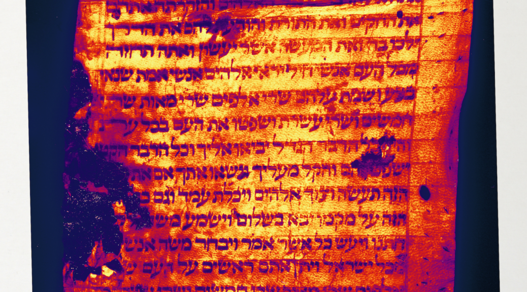
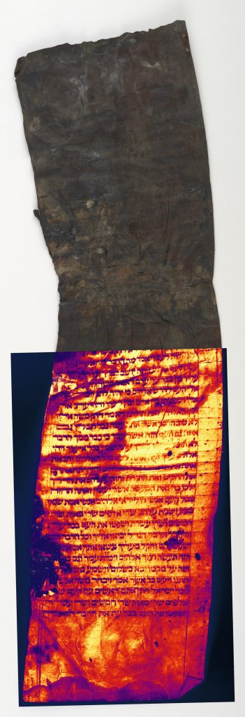
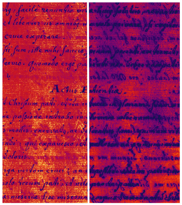
Below are two stacked images of Ashkar MS 16 (from the Rubenstein Library).  The top half of each image is the manuscript under natural light, and the bottom are the results of Multispectral imaging and processing.  We tend to post black and white MSI images most often as they are generally the most legible, however our MSI software can produce a lot of wild color variations!  The orange one below seemed the most appropriate for a hot NC July afternoon like today.  More processing details are included in the image captions below – enjoy!

The text of this manuscript above was revealed primarily with the IR narrowband light at 780 nm.
This image was created using Teem, a tool used to process and visualize scientific raster data. This specific image is the result of flatfielding each wavelength image and arranging them in wavelength order to produce a vector for each pixel. The infinity norm is computed for each vector to produce a scalar value for each pixel which is then histogram-equalized and assigned a color by a color-mapping function.

Announcing Multispectral Imaging Service, version 1

As regular Bitstreams readers know, a cross departmental team within Duke University Libraries has been exploring Multispectral Imaging and its potential to make Duke collections more accessible to researchers in the Duke scholarly community and beyond since 2015. After spending 2017 developing MSI workflows, building expertise, writing documentation, and responding to experimental imaging requests, we are now ready to unveil the first version of Duke University Libraries MSI service for researchers!

Our first service model version accommodates small requests that are not urgent. The MSI team wants to partner with researchers to facilitate their requests as well as hear feedback about our current service and any other needs for MSI. We are offering MSI services for free for the next few months, but will institute a fee structure this Summer.

The service breaks down into 4 general steps:

  • First, researchers submit a request for MSI services using a webform. The form prompts requesters to share their research question and details about what they want imaged. We also want to know where researchers are from, as we are expecting both Duke and non-Duke affiliated patrons.
  • Second, the MSI team will review all requests, as MSI is not the ideal imaging solution for all materials and research questions. Requests that will not benefit from MSI will not be approved.
  • Third, we schedule approved requests for imaging and processing. We plan to conduct 1 imaging and processing day per month, so it may take several weeks to a month for approved requests to make it though our full process.
  • Fourth, we deliver the processed files to our patrons along with a report that details the imaging and processing procedures and outcomes.

Please note the following:
We are currently only imaging Duke University Library holdings.
We are limiting requests to 1-3 individual items or 1-3 pages within a bound item (which is the number of items we can generally image and process in 1 day).
Allow 2-4 weeks for vetting and up to a month for imaging.

If you are interested in requesting MSI services, but your needs do not fit the service described here, we still want to hear from you! Please do not hesitate to fill out our researcher request form to get the process started, or contact Susan Ivey directly.

If you want to learn more about MSI, check out the recent talk we gave at the Friday Visualization Forum on February 23.

Institutional multispectral imaging (MSI) survey results

Over the last year and a half, we’ve blogged quite a bit about our exploration of multispectral imaging (MSI) here at Duke Libraries. We’ve written about the hardware and software that we acquired in 2016 and about our collaboration and training to help us learn how to use this new equipment. We’ve shared examples from our experiments with MSI on various material and ink types and talked about how MSI can help inform conservation treatment and help uncover faded or hidden text.

Periodically, we’ve also mentioned our Duke MSI group. This group is a cross-departmental team of individuals that each bring to the table critical expertise to ensure that our MSI projects run smoothly and effectively. This past year, our team has created various best practices and workflows, tackled equipment and software hurdles, determined current resource capacity, imaged a number of materials from Duke’s collections, and documented data modeling scenarios. We’re really excited about and proud of the work that we’ve accomplished together.

Many times, though, we’ve talked about how helpful it would be to talk with other institutions that are doing MSI, particularly cultural heritage institutions and academic libraries, in order to learn how they develop their skills and organize their services. Luckily, the University of Manchester, an institution that has the same MSI system as ours, expressed interest in collaborating with us to our shared vendor R. B. Toth Associates. We had a very informative conference call with them in May 2017, during which we discussed their program, services, and technical details. We also shared documentation after the call, which has helped us refine our project request procedures and our deliverables that we provide to the requesters at the end of projects.

Since that collaboration proved so beneficial, we thought it would be helpful to reach out to other institutions that undertake MSI projects. Though it is difficult to determine exact numbers online, it is clear that very few cultural heritage organizations and academic libraries host their own MSI system. Therefore, we teamed up with R. B. Toth Associates again to determine what other institutions they’d worked with that may be interested in collaborating. They shared six institutions worldwide, and we created a brief online survey about the topics that we’d discussed with the University of Manchester, which included organization and staffing, time devoted to MSI, prioritization, project deliverables, and data modeling and access. Five of the six institutions thought that the survey applied to their current MSI setup and therefore completed the survey, and all six expressed some level of interest in future collaboration. The institutions that completed the survey are the Library of Congress, the Museums of New Mexico-Conservation, the University of Pennsylvania Libraries, KU Leuven Libraries, and the University of Copenhagen.

Out of the institutions that we’ve spoken with or surveyed (six in total, including the University of Manchester), we can determine that three have permanent MSI systems (the Library of Congress, the Museums of New Mexico, and University of Manchester). One institution’s survey results were unclear about owning its own system, and the other two institutions noted that they use MSI systems on an ad-hoc, project by project basis. One of those institutions indicated that they are in the initial stages of considering the purchase of a permanent system, and the institution that declined taking the survey stated that it was because they did not own their own system due to costs. Some institutions charge a fee for MSI projects, while others don’t. One institution that currently does not charge expressed interest in creating a cost recovery model.

In terms of the institutions that own a permanent MSI system, the Library of Congress is the only that has one full time employee devoted solely to MSI. The Museums of New Mexico have one employee that serves as the main PI for MSI, though the entire Conservation department is trained on the equipment and software. Their department devotes approximately 20 hours per week to MSI depending on the project. The University of Manchester has two imagers that devote one day per month to imaging, and process intermittently. The other institutions without permanent systems use a combination of trained staff, scholars, imaging scientists, object handlers and conservators, metadata and data managers, imaging technicians, and project managers based on specific project needs.

It appears that most institutions image their own collections for MSI projects, though one respondent indicated that they also partner with other conservation institutions for their research needs. Prioritization processes for MSI projects vary institution to institution. The Library of Congress is the exception, because that department solely does MSI projects, and therefore requests do not need to be prioritized with other digitization requests. The Museums of New Mexico prioritize on a first come, first serve basis, as long as the project is a good fit for MSI. The University of Manchester also has a pre-project process with conservation staff to determine if the project is a good fit for MSI. KU Leuven Libraries prioritizes based on scope, indicating the priority is documentary heritage with a strong focus on illuminated manuscripts, but will service other research if time permits. Additional prioritization strategies include availability of trained staff and scholars.

The institutions have similar goals for MSI projects based on whether a project is internal (for conservation or acquisition purposes) or external (research requested). These include pure documentation, establishing a condition baseline, forensic examination and reconstruction, pre-purchase examination, collecting spectral responses for preservation, revealing undertext for transcription, and revealing undertext for text identification.

Deliverables and preservation copies vary by institution as well. Three of the institutions always include some type of a report with the files. KU Leuven provides multiple reports, including a technical report about infrastructure, an analysis report, and an imaging methodology. Two of the institutions provide the raw and processed images, two provide only the processed images (as jpegs or tiffs), and two state that the deliverables vary based on project. The University of Pennsylvania is the only institution that noted that it provides metadata, which often include transcriptions or descriptive and structural metadata and is packaged in a preservation-ready package. One respondent replied that they retain raw data offline, but place processed captures into a digital asset management system (presumably for online access), and another noted that some projects have arranged preservation of their data packages through third parties that offer dark or accessible digital repositories. Every institution retains some or all of their MSI data internally.

This is just a brief summary of what we learned through this process—we were lucky to receive a lot of data and comments! We are very pleased with and thankful for their willingness to share. As noted earlier, all have expressed an interest in some level of future collaboration, so we’re hopeful to build a network of institutions that can provide feedback and advice to one another for future MSI needs. To all of our fellow MSI institutions, we thank you again for your participation! Please let me know if I misrepresented any of your information. We received a lot of great qualitative responses, and though I did my best to communicate those correctly, I may have misunderstood some responses.


Multispectral Imaging and Thermofaxes

Duke University Libraries has successfully used multispectral imaging to make ancient manuscripts more readable, but the same technology can also be employed on modern collection materials.  Over the summer, our multispectral imaging working group teamed up with Rubenstein Library staff to investigate whether advanced imaging techniques could be used to make certain faded photocopies more legible. Duke’s archival collections are filled with copies of documents made using a wide range of printing techniques. Some print processes which were popular before the proliferation of “laser” printing (electrostatic prints) can become badly and irreversibly faded.  The Thermo-Fax was a specific type of copying process, introduced by 3M in 1950, but has become a common proprietary eponym for this whole category of collection materials.

Robert Hill Papers, Electrolytic print under normal illumination

When confronted with a badly faded photocopy, library staff have traditionally resorted to one preservation tactic: create a more stable reproduction of the document on high quality paper. While the original print will continue to fade over time, the “preservation photocopy” should remain just as legible. In some cases, the decision may be made to retain just the preservation copy and deaccession the original. If the document is completely illegible, however, a preservation photocopy won’t be effective and the original may not be retained at all; it’s difficult to determine the value of a letter that can’t be read. If alternative imaging techniques could be used to produce a better reproduction, it might dramatically change how we assess and work with faded documents.

Meghan Lyon, Manuscript Processing Archivist at Rubenstein Library Technical Services, helped to identify several examples of early photocopies from various collections. Our first trial was with a print, probably dating from the 1970s, from the Robert A. Hill Collection of the Marcus Garvey and Universal Negro Improvement Association Papers Project Records (see image above). This collection contains many reproductions of newspaper articles from microfilm. After multispectral imaging and software processing , we achieved these results:

Robert HIll Papers, PCA with False Color

The above image is more legible than under regular illumination, but really not the unequivocal win for which we had hoped. Research about machines that would print copies from microfilm suggested that this type of print, an electrolytic print process sometimes called “Photoconductography”, may not be the best candidate. The image is generated from metallic silver, rather than a dye. Therefore, this item likely became damaged from a water event, rather than fading. We went back to our friends in Technical Services for more examples.

Radio Haiti Collection during processing

The next candidate for imaging came from the Radio Haiti Archives. This collection contains more modern prints (from the 1990s), which are completely faded. Our test document (pictured below) almost looks like a blank sheet of paper. The only visible markings are from a ballpoint pen. Based upon descriptions of paper types and qualities of this object, we identified this as a form of direct thermal printing.

Radio Haiti, Direct thermal print under normal illumination

The paper used in this type of direct thermal printing is impregnated with a colorless dye precursor and coupler system. Thermoplastic material separates the two ingredients. When exposed to heat, the separator melts and the precursor and coupler react to form the colored print. Dyes are notoriously unstable over time, so it is not uncommon for thermal prints to completely disappear. Our document exhibits other interesting properties. For example, the faded text is visible when the page is held up to a light source. We often take transmitted light photos as part of conservation treatment documentation, so I imaged the object on top of a lightbox in a camera copy stand. This technique was pretty successful in delivering a readable image.

Radio Haiti, Transmitted light imaging

It is helpful to know that a low-tech method for reading or imaging this faded documents can be employed. It is easy to imagine library staff using a simple lightbox to identify, assess, or arrange a large collection of prints like this one.

Multispectral imaging of the letter yielded much better results. Interestingly, additional visual noise was introduced from fingerprints and hand marks on the page.

Radio Haiti, PCA with false color

Looking through the image stack, it appears the text is most legible under ultraviolet light (370nm) with a neutral filter in front of the camera.

Radio Haiti, 370nm

This result is actually quite promising when we think about scaling up an imaging project for an entire collection. Creating a full 18 image stack and processing those images takes a great deal of time; however, if we know that one frequency of light and filter combination is effective, we could easily set the imaging equipment to operate more like a normal copy stand.  In this way, we could bring the requisite imaging time and storage costs closer to those of our regular imaging projects.

Many modern print types have inherent vices that cause them to fade rapidly and large collections of them are common in library archives. Advanced imaging techniques, like multispectral imaging, potentially offer opportunities to identify, arrange, and preserve volatile or unreadable prints. Creating a full image stack may not be possible for every item in a large collection, but this investigation illustrates that full MSI may not be required to achieve the needed results.

More MSI Fun: Experimenting with iron gall ink

Submitted on behalf of Erin Hammeke

For conservators, one of the aspects of having the MSI system that excites us most is being able to visualize and document the effects of the treatments that we perform. Although we are still learning the ropes with our new system, we had a recent opportunity to image some iron gall ink documents. Iron gall ink is common historic ink that reacts with moisture in the environment to form acidic complexes that spread and sink into the paper, weakening the paper and, in some cases, leaving holes and losses. This iron gall ink degradation can be better visualized with MSI, since the beginning stage, haloing, is not always visible under normal illumination. Look here for more information on iron gall ink damage and here for using MSI to document iron gall ink condition and treatment. We also illustrated the haloing effect of iron gall ink damage using MSI on Jantz MS #124 in a previous post.

Recently, DUL conservators experimented with treating some discarded iron gall ink manuscripts with a chemical treatment that aims to arrest the ink’s degradation. This treatment requires submerging the manuscripts in a calcium phytate solution – a chemical that bonds with free iron (II) ions, stabilizing the ink and preventing it from corroding further. The document is then rinsed in a water bath and an alkaline reserve is applied. Resizing with gelatin is another common step, but we did resize our test manuscripts.

Since these were discarded test material, we were able to cut the manuscripts in two and only treat one half. Imaging the manuscript with MSI revealed some notable findings.

Most of the treated papers now appear lighter and brighter under normal illumination because they have been washed. However, the untreated halves exhibited pronounced UV induced visible fluorescence around the 488 nm range and the treated halves did not. We believe this difference likely has to do with washing the paper substrate and rinsing out degradation products or perhaps paper size that may exhibit fluorescence at this wavelength. We were happy to see that for a treatment that targets the ink, there was very little noticeable difference in the appearance of the inks between untreated and treated portions of the test manuscript. There was some reduction in the “ink sink” (ink visible from the opposite side of the manuscript) and a very slight softness to the edges of the ink in the treated sample, but these changes were very minimal. We look forward to imaging more of our test manuscripts in the future and seeing what else we can learn from them.

______

Want to learn even more about MSI at DUL?

Infrastructure and Multispectral Imaging in the Library

As we continue to work on our “standard” full color digitization projects such as Section A and the William Gedney Photograph Collection, both of which are multiyear projects, we are still hard at work with a variety of things related to Multispectral Imaging (MSI).  We have been writing documentation and posting it to our Knowledgebase, building tools to track MSI requests and establishing a dedicated storage space for MSI image stacks.  Below are some high-level details about these things and the kinks we are ironing out of the MSI process.  As with any new venture, it can be messy in the beginning and tedious to put all the details in order but in the end it’s worth it.

MSI Knowledge Base

We established a knowledge base for documents related to MSI that cover a wide variety of subjects:  How-To articles, to do lists, templates, notes taken during imaging sessions, technical support issues and more.  These documents will help us develop sound guidelines and workflows which in turn will make our work in this area more consistent, efficient and productive.

Dedicated storage space

Working with other IT staff, a new server space has been established specifically for MSI.  This is such a relief because, as we began testing the system in the early days, we didn’t have a dedicated space for storing the MSI image stacks and most of our established spaces were permissions restricted, preventing our large MSI group from using it.  On top of this we didn’t have any file management strategies in place for MSI.  This made for some messy file management. From our first demo, initial testing and eventual purchase of the system, we used a variety of storage spaces and a number of folder structures as we learned the system.  We used our shared Library server, the Digital Production Center’s production server, Box and Google Drive.  Files were all over the place!  What a mess!  In our new dedicated space, we have established standard folder structures and file management strategies and store all of our MSI image stacks in one place now.  Whew!

The Request Queue

In the beginning, once the MSI system was up and running, our group had a brainstorming session to identify a variety of material that we could use to test with and hone our skills in using the new system.  Initially this queue was a bulleted list in Basecamp identifying an item.  As we worked through the list it would sometimes be confusing as to what had already been done and what item was next.  This process became more cumbersome because multiple people were working through the list at the same time, both on capture and processing, with no specific reporting mechanism to track who was doing what.  We have recently built an MSI Request Queue that tracks items to be captured in a more straightforward, clear manner.  We have included title, barcode and item information along with the research question to be answered, it priority level, due date, requester information and internal contact information.  The MSI group will use this queue for a few weeks then tweak it as necessary.  No more confusion.

The Processing Queue

As described in a previous post, capturing with MSI produces lots of image stacks that contain lots of files.  On average, capturing one page can produce 6 image stacks totaling 364 images.  There are 6 different stages of conversion/processing that the image stack goes through before it might be considered “done”, and the fact that everyone on the MSI team has other job responsibilities makes it difficult to carve out a large enough block of time to convert and process the image stacks through all of the stages.  This made it difficult to know what items had been completely processed or not.  We have recently built an MSI Processing Queue that tracks what stage of processing each item is in.  We have included root file names, flat field information, PPI and a column for each phase of processing to indicate whether or not an image stack has passed through a phase.  As with the Request Queue, the MSI group will use this queue for a few weeks then tweak it as necessary.  No more confusion.

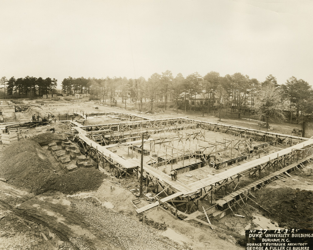
Duke University East Campus Progress Picture #27

As with most blog posts, the progress described above has been boiled down and simplified as to not bore you to death, but this is a fair amount of work nonetheless.  Having dedicated storage and a standardized folder structure simplifies the management of lots of files and puts them in a predictable structure.  Streamlining the Request Queue establishes a clear path of work and provides enough information about the request in order to move forward with a clear goal in mind.  Developing a Processing Queue that provides a snapshot of the state of processing across multiple requests and provides enough information so that any staff member familiar with our MSI process can complete a request.  Establishing a knowledge base to document our workflows and guidelines ties everything together in an organized and searchable manner making it easier to find information about established procedures and troubleshoot technical problems.

It is important to put this infrastructure in place and build a strong foundation for Multispectral Imaging at the Library so it will scale in the future.  This is only the beginning!

_______________________

Want to learn even more about MSI at DUL?


Multispectral Imaging: What’s it good for?

At the beginning of March, the multispectral imaging working group presented details about the imaging system and the group’s progress so far to other library staff at a First Wednesday event. Representatives from Conservation Services, Data and Visualization Services, the Digital Production Center, the Duke Collaboratory for Classics Computing (DC3) and the Rubenstein Library each shared their involvement and interest in the imaging technology. Our presentation attempted to answer some basic questions about how the equipment works and how it can be used to benefit the scholarly community. You can view a video of that presentation here

Some of the images we have already shared illustrate a basic benefit or goal of spectral imaging for books and manuscripts: making obscured text visible. But what else can this technology tell us about the objects in our collections? As a library conservator, I am very interested in the ways that this technology can provide more information about the composition and condition of objects, as well as inform conservation treatment decisions and document their efficacy.

Conservators and conservation scientists have been using spectral imaging to help distinguish between and to characterize materials for some time. For example, pigments, adhesives, or coatings may appear very differently under ultraviolet or infrared radiation. Many labs have the equipment to image under a few wavelengths of light, but our new imaging system allows us to capture at a much broader range of wavelengths and compare them in an image stack.

Adhesive samples under visible and UV light.
(Photo credit Art Conservation Department, SUNY Buffalo State)

Spectral imaging  can help to identify the materials used to make or repair an object by the way they react under different light sources. Correct identification of components is important in making the best conservation treatment decisions and might also be used to establish the relative age of a particular object or to verify its authenticity.  While spectral imaging offers the promise of providing a non-destructive tool for identification, it does have limitations and other analytical techniques may be required.

Pigment and dye-based inks under visible and infrared light.
(Photo credit Image Permanence Institute)

Multispectral imaging offers new opportunities to evaluate and document the condition of objects within our collections. Previous repairs may be so well executed or intentionally obscured that the location or extent of the repair is not obvious under visible light. Areas of paper or parchment that have been replaced or have added reinforcements (such as linings) may appear different from the original when viewed under UV radiation. Spectral imaging can provide better visual documentation of the degradation of inks ( see image below) or damage from mold or water that is not apparent under visible light.

Iron gall ink degredation. Jantz MS#124, Rubenstein Library
(Jantz MS#124, Rubenstein Library)

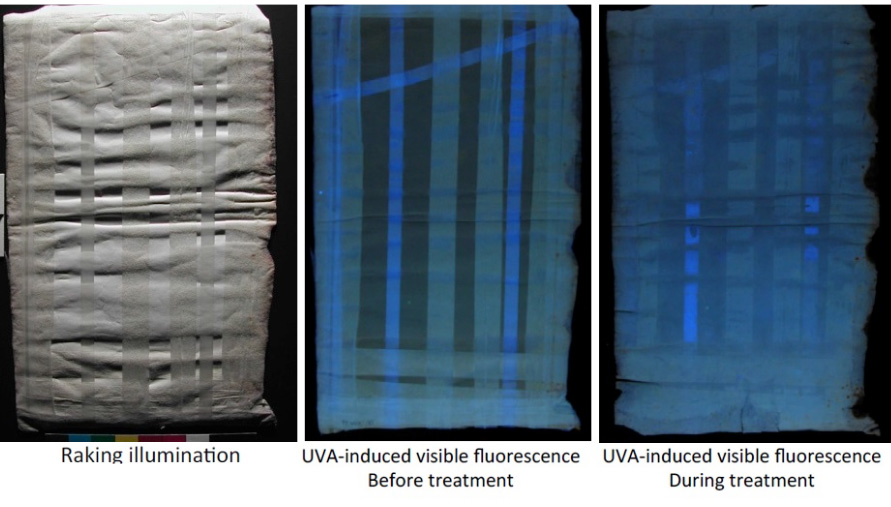
This imaging equipment provides opportunities for better measuring the effectiveness of the treatments that conservators perform in-house. For example, a common treatment that we perform in our lab is the removal of pressure sensitive tape repairs from paper documents using solvents. Spectral imaging before, during, and after treatment could document the effectiveness of the solvents or other employed techniques in removing the tape carrier and adhesive from the paper.

Tape removal before and during treatment under visible and UV light.
(Photo credit Art Conservation Department, SUNY Buffalo State)

Staff from the Conservation Services department have a long history of participating in the library’s digitization program in order to ensure the safety of fragile collection materials. Our department will continue to serve in this capacity for special collections objects undergoing multispectral imaging to answer specific research questions; however, we are also excited to employ this same technology to better care for the cultural heritage within our collections.

______

Want to learn even more about MSI at DUL?


Multispectral Imaging Through Collaboration

I am sure you have all been following the Library’s exploration into Multispectral Imaging (MSI) here on Bitstreams, Preservation Underground and the News & Observer.  Previous posts have detailed our collaboration with R.B. Toth Associates and the Duke Eye Center, the basic process and equipment, and the wide range of departments that could benefit from MSI.  In early December of last year (that sounds like it was so long ago!), we finished readying the room for MSI capture, installed the equipment, and went to MSI boot camp.

Obligatory before and after shot. In the bottom image, the new MSI system is in the background on the left with the full spectrum system that we have been using for years on the right. Other additions to the room are blackout curtains, neutral gray walls and black ceiling tiles all to control light spill between the two camera systems. Full spectrum overhead lighting and a new tile floor were installed which is standard for an imaging lab in the Library.

Well, boot camp came to us. Meghan Wilson, an independent contractor who has worked with R.B. Toth Associates for many years, started our training with an overview of the equipment and the basic science behind it. She covered the different lighting schemes and when they should be used.  She explained MSI applications for identifying resins, adhesives and pigments and how to use UV lighting and filters to expose obscured text.   We quickly went from talking to doing.  As with any training session worth its salt, things went awry right off the bat (not Meghan’s fault).  We had powered up the equipment but the camera would not communicate with the software and the lights would not fire when the shutter was triggered.  This was actually a good experience because we had to troubleshoot on the spot and figure out what was going on together as a team.  It turns out that there are six different pieces of equipment that have to be powered-up in a specific sequence in order for the system to communicate properly (tee up Apollo 13 soundtrack). Once we got the system up and running we took turns driving the software and hardware to capture a number of items that we had pre-selected.  This is an involved process that produces a bunch of files that eventually produce an image stack that can be manipulated using specialized software.  When it’s all said and done, files have been converted, cleaned, flattened, manipulated and variations produced that are somewhere in the neighborhood of 300 files. Whoa!

This is not your parents’ point and shoot—not the room, the lights, the curtains, the hardware, the software, the pricetag, none of it. But it is different in another more important way too. This process is team-driven and interdisciplinary. Our R&D working group is diverse and includes representatives from the following library departments.

  • The Digital Production Center (DPC) has expertise in high-end, full spectrum imaging for cultural heritage institutions along with a deep knowledge of the camera and lighting systems involved in MSI, file storage, naming and management of large sets of files with complex relationships.
  • The Duke Collaboratory for Classics Computing (DC3) offers a scholarly and research perspective on papyri, manuscripts, etc., as well as  experience with MSI and other imaging modalities
  • The Conservation Lab brings expertise in the Libraries’ collections and a deep understanding of the materiality and history of the objects we are imaging.
  • Duke Libraries’ Data Visualization Services (DVS) has expertise in the processing and display of complex data.
  • The Rubenstein Library’s Collection Development brings a deep understanding of the collections, provenance and history of materials, and valuable contacts with researchers near and far.

To get the most out of MSI we need all of those skills and perspectives. What MSI really offers is the ability to ask—and we hope answer—strings of good questions. Is there ink beneath that paste-down or paint? Is this a palimpsest? What text is obscured by that stain or fire-damage or water damage? Can we recover it without having to intervene physically? What does the ‘invisible’ text say and what if anything does this tell us about the object’s history? Is the reflectance signature of the ink compatible with the proposed date or provenance of the object? That’s just for starters. But you can see how even framing the right question requires a range of perspectives; we have to understand what kinds of properties MSI is likely to illuminate, what kinds of questions the material objects themselves suggest or demand, what the historical and scholarly stakes are, what the wider implications for our and others’ collections are, and how best to facilitate human interface with the data that we collect. No single person on the team commands all of this.

Working in any large group can be a challenge. But when it all comes together, it is worth it. Below is a page from Jantz 723, one processed as a black and white image and the other a Principal Component Analysis produced by the MSI capture and processed using ImageJ and a set of tools created by Bill Christens-Barry of R.B. Toth Associates with false color applied using Photoshop. Using MSI we were able to better reveal this watermark which had previously been obscured.

Jantz 723

I think we feel like 16-year-old kids with newly minted drivers’ licenses who have never driven a car on the highway or out of town. A whole new world has just opened up to us, and we are really excited and a little apprehensive!

What now?

Practice, experiment, document, refine. Over the next 12 (16? 18) months we will work together to hone our collective skills, driving the system, deepening our understanding of the scholarly, conservation, and curatorial use-cases for the technology, optimizing workflow, documenting best practices, getting a firm grip on scale, pace, and cost of what we can do. The team will assemble monthly, practice what we have learned, and lean on each other’s expertise to develop a solid workflow that includes the right expertise at the right time.  We will select a wide variety of materials so that we can develop a feel for how far we can push the system and what we can expect day to day. During all of this practice, workflows, guidelines, policies and expectations will come into sharper focus.

As you can tell from the above, we are going to learn a lot over the coming months.  We plan to share what we learn via regular posts here and elsewhere.  Although we are not prepared yet to offer MSI as a standard library service, we are interested to hear your suggestions for Duke Library collection items that may benefit from MSI imaging.  We have a long queue of items that we would like to shoot, and are excited to add more research questions, use cases, and new opportunities to push our skills forward.   To suggest materials, contact Molly Bragg, Digital Collections Program Manager (molly.bragg at Duke.edu), Joshua Sosin, Associate Professor in Classical Studies & History (jds15 at Duke.edu) or Curator of Collections (andrew.armacost at Duke.edu).

Want to learn even more about MSI at DUL?