Tag Archives: history of medicine

Lecture: The History and Legacy of Ivory Anatomical Manikins

Date: Tuesday, October 25, 2016
Time: 4:00 p.m.
Location: Rubenstein Library Room 153 (Holsti-Anderson Family Assembly Room)

Cali Buckley
Cali Buckley

Join the Trent History of Medicine Lecture Series for our next lecture by Cali Buckley on “The History and Legacy of Ivory Anatomical Manikins.” Ivory anatomical models comprise a little-known set of objects that were popular with male doctors of the late 17th- and 18th-centuries. Their narrative is currently being revised in light of a history of questionable assumptions. Though small and largely inaccurate, the story of anatomical manikins reveals how the politics of medicine impresses meaning on medical objects – often transcending the needs of the scientific community. Ms. Buckley will present on her current hypotheses as well as the process by which medical objects can be examined according to social history, connoisseurship, and material culture.

Cali Buckley is a Ph.D. candidate in art history at Penn State University. She received a Fulbright U.S. Student Award that allowed her to spend the 2015–16 academic year in Germany working on her dissertation, “Early Modern Anatomical Models and the Control of Women’s Medicine.”

The talk will be held in the Holsti-Anderson Family Assembly Room, Room 153, of the Rubenstein Library at Duke University. All are welcome to attend.  Sponsored by the History of Medicine Collections.

Measuring the Children of the Corn

“The babies are entered like any other exhibit at an agricultural fair. . . . They are examined by judges, just as live stock [sic], grain or apples are examined. . . . The result is bound to be – not prettier babies, – but better babies at each year’s fair, – a stronger, healthier race of people on the farms, in small towns and in the state.”

This excerpt from a Better Babies Bureau circular, from the papers of Victor Bassett, contains several templates for Better Baby Contest advertisements. Popular at local fairs in the early 20th century, Better Baby Contests presented a lighthearted way to challenge infant mortality and promote fitter populations. However, they also reveal governmental eugenic efforts to objectively quantify and thus improve American health.

Announcement for Better Babies Contest.

Examiners judged children under five years old on their measurements and proportions, mental and developmental states, vision and hearing, and physical development. These tests set government-determined averages as the standard by encouraging families to “produce” children who met or exceeded these ideals. Additionally, they served to cement the public health as existing in the realm of scientific medicine and the government, rather than in the home. These kinds of contests illustrate the complex relationship between eugenics, popular movements, and public health.

Thanks to a generous History of Medicine travel grant, my visit to the Rubenstein Rare Book & Manuscript Library allowed me to conduct research to support my dissertation, “Measuring Health: The United States Sanitary Commission, Statistics, and American Public Health in the Nineteenth Century.” I examine the statistical work of the United States Sanitary Commission (USSC).

During the American Civil War, the USSC attempted to improve the health of the Union Army. The leaders of this organization understood their work as forwarding ideas about preventative medicine and improving sanitation. Yet with the entirety of the Union army at their disposal, the USSC also inspected and measured over one million soldiers and sailors. These records include tabulations not simply of height and weight, but also the distance between a man’s eyes, the size of his head, and angles of his face. The statisticians presented their findings in groups divided by race and education level, and, while they provided limited interpretation of these numbers, they were made available to the broader scientific and anthropologic communities. It was in their hands that these numbers defined medical standards for Americans and shaped the nature of American public health.

My project explores why these statistics were collected and how they were used, and, more broadly, the Commission’s public health legacy. By using these statistics as a starting point, I explore the ties between the USSC and changes in public health, and how research from a 19th century organization continues to impact later public health issues. These Better Baby Contests represent the Commission’s legacy of measuring the quality and usefulness of a human being and of using governmental authority to establish scientific authority.

Post contributed by Sara Kern, a Ph.D. candidate in History & Women’s, Gender, and Sexuality Studies at Penn State University.

Photographic Research on Obstetric and Gynecological Instruments

Auvard Weighted Speculum from the L. M. Draper Collection..
Auvard Weighted Speculum from the L. M. Draper Collection.

With generous assistance from the History of Medicine travel grant, I traveled to Duke University to view and photograph historical obstetric and gynecological tools housed in Duke’s History of Medicine Collections at the Rubenstein Rare Book & Manuscript Library.  There I viewed various artifact collections donated by practicing regional doctors, including the L. M. Draper Collection, the George D. & Evelyn Wilbanks Collection, and several anonymous collections.  I also viewed anatomical lift-the-flap guide books, lift-the-flap anatomical fugitive sheets and the Trent Collection of Ivory Anatomical Manikins, all of which were used to teach medical procedures, including delivery.

Having access to Duke’s collection was an incredible experience.  I treated it like a short artist residency.  I set up my lights, a pop-up tent, my camera and a tripod in a study room within the library.  Every morning, a cart was wheeled in with OB/GYN tools, anatomy text books and glass slides.  It was exciting (and a little nerve-wracking), opening up boxes and not knowing their contents.  For some items, I felt I was discovering the files for the first time.  In a way I was: besides the archivists who received and catalogued them, some of the items had never been requested.  I often felt as though I were in the medical field—donning nitrile gloves, carefully removing the items from their boxes, gently lying them down on the fabric of my pop-up lighting tent, careful not to harm them in any way.  I found myself photographing them as abstractions or as jewelry, a style of cataloguing unlike other projects I have photographed.

Smellie-Style Obstetrical Forceps from the George & Evelyn Wilbanks Collection.

My work focuses on historical and contemporary women’s lives and I am particularly interested in the past’s technology and how it relates to today.  I have previously done photographic projects on antique vibrators, social media and the practice of keeping a commonplace book and with this project, the history of labor and delivery technology.  While the process of getting pregnant has changed with IVF and the location of delivery may have changed, the actual process of delivery has not changed.  Although American society emphasizes new products & experiences, and the medical world uses recent technology & procedures, women continue to deliver only one of two ways—vaginally or via Cesarean section.  Prior to my arrival at Duke, I assumed the tools used in labor and delivery were harmful to the infants and delivering women.  I also wondered how deadly labor actually was—in fictionalized accounts in both books and screen, no female who delivered a newborn ever lived, and seldom the child.  I expected antique tools to be brutal and different in appearance than today.  It surprised me that many of the tools I photographed resembled contemporary tools, only with time’s effect through rust or evident aging.

My research at Duke is the beginning of both my project and into further research on the history of the OB/GYN tools and their uses.  Although in its early stages, I plan to study these tools’ history, as well as their use & influence today.  The final images may be printed as slides, emulating turn of the twentieth century magic-lantern plates or late-twentieth century educational slide shows.  Whatever form these images take, I was particularly inspired by the anatomical lift-the flap books & broad sides and will create an artist book influenced by these interactive educational guides.  I look forward to sharing future developments of this project. Thank you to everyone at Duke University’s Rubenstein Library for their assistance during my stay.

Post contributed by History of Medicine Travel Grant recipient Lindsey Beal. Beal is a photo-based artist and professor in Providence, Rhode Island.  Her work and further information can be found at lindseybeal.com.

Trent Lecture on the Brooklyn Interns Hazing Episodes: March 7th

Date: Monday, March 7, 2016
Time: 5:30 PM
Location: Holsti-Anderson Family Assembly Room, Room 153, Rubenstein Library
Contact: Rachel Ingold, rachel.ingold@duke.edu, (919) 684-8549

Dr. Ed Halperin, M.D., M.A.Please join the History of Medicine Collections for our next Trent History of Medicine Lecture Series event. Dr. Ed Halperin, M.D., M.A., will present “This is a Christian institution and we will tolerate no Jews here”:  The Brooklyn Interns Hazing Episodes.

Anti-semitism in U.S. medical education rarely flared into acts of violence, except in Brooklyn. Presenting the results of recently completed research, Dr. Halperin will describe the assaults on the Jewish interns of Kings County Hospital in 1916 and 1927 and the implications of these assaults for the contemporary debate on immigration and higher education.

Dr. Halperin is Chancellor  and Chief Executive Officer at New York Medical College and Professor of Radiation Oncology, Pediatrics, and History as well as Provost for Biomedical Affairs at Touro College.

This event is open to the public.

Post contributed by Rachel Ingold, Curator of the History of Medicine Collections.

Upcoming Talk: Scientists, Midwives, & Healers in the Lisa Unger Baskin Collection

Date: Tuesday, February 9, 2016
Time: 4:00 PM
Location: Holsti-Anderson Family Assembly Room, Rubenstein Library Room 153
Contact: Laura Micham, laura.m@duke.edu
RVSP (optional) via Facebook

Maria Sibylla Merian. De europische insecten. Tot Amsterdam: by J.F. Bernard, [1730].
Maria Sibylla Merian. De europische insecten. Tot
Amsterdam: by J.F. Bernard, [1730].
Join the staff of the Bingham Center as Duke History Professor Thomas Robisheaux gives a lecture on the Lisa Unger Baskin Collection, highlighting his use of works by naturalist and artist Maria Sibylla Merian. The lecture is free and open to the public; light refreshments will be served.

In celebration of:

Heralding the Way to a New World: Exploring Women in Science and Medicine through the Lisa Unger Baskin Collection

On display in the Michael and Karen Stone Family Gallery from January 20th to April 1st, 2016

From the first entomologist to capture the stages of metamorphosis of the butterfly (1705) to the author who published the first comprehensive volume on contraception (1923), the women in this exhibit were pioneers in science and medicine. Whether self-trained or classically educated, they not only made groundbreaking contributions to their fields, but also helped open the way for future generations to follow in their footsteps. Despite their accomplishments, most of these women remain overlooked or under-recognized.

This exhibition highlights the stories of seven revolutionary women in science and medicine and celebrates the arrival of the Lisa Unger Baskin Collection, from which these materials were selected.

Frederik Ruysch’s Anatomical Art

Post contributed by Amelia Holmes, Josiah Charles Trent Intern for the History of Medicine Collections.

Anatomical specimens emerged as an art form near the end of the seventeenth century. Although they may seem morbid today, at the time of creation, they were viewed as striking a balance between the scientific and the artistic. They served to educate people on human anatomy as well as to remind them of the fleeting nature of life.

Illustration from Opera Omnia Anatomico-Medico-Chirurgica, ca. 1737.

One of the more notable creators of anatomical art is Frederik Ruysch, a Dutch botanist and anatomist who lived from 1638 to 1731—an impressive 93 years in a time when many died young. A capable researcher, Ruysch was the first to describe bronchial blood vessels, the vascular plexus of the heart, and the valves of the lymphatics. However, his real interest lay in anatomical preparations, and he has been described by a recent biographer as “probably the most skilled and knowledgeable preparator in the history of anatomy” (Gould, p. 20). Ruysch served as the chief instructor to midwives and the “legal doctor” to the court of Amsterdam. Through these positions, he had easy (and legal) access to the bodies of stillborns and dead babies.

The preparations were initially created to use in his classes, but they eventually gained an interest from the public. To showcase his vast collection (he created more than 2,000 from 1665 to 1717 alone), he opened his own cabinet of curiosities to the public, which for many marked the first time they were able to see human internal organs. The collection was also noteworthy because of the lengths to which Ruysch went in an effort to make the specimens appear more natural. For example, embalmed children were clothed or held bouquets of preserved flowers. In 1717, Peter the Great, who was an admirer of Ruysch, purchased the entire cabinet of curiosities for 30,000 guilders. The collection was then shipped to St. Petersburg, and along with the cabinet of curiosities formed by Albertus Seba, they became the core of the Kunstkammer—the Academy of Sciences of Russia’s first public museum.

Although a number of Ruysch’s wet preparations still exist today (a fact which he would find unsurprising), none of his dry specimens have been located. He used fetal skeletons and other body parts to create multi-specimen scenes. These scenes served as the centerpieces for each of the literal cabinets within the rooms of his museum. As Gould points out, these tableaux were focused on allegorical themes such as death and the transience of life. The small skeletons are decorated with symbols of death and short life: mayflies rest in hands, skulls weep into handkerchiefs made of mesentery, and snakes made of intestine wine their way through bones. Today, these still-life scenes exist for us only through second-hand descriptions and, fortunately, through a number of engravings.

Fortunately for those interested in seeing these illustrations up close, the History of Medicine Collections has two volumes from the multi-volume Opera omnia anatomico-medico-chirurgica. On October 29, from 2-4 pm, they will be on display as part of Screamfest in the Holsti-Anderson Family Assembly Room.

Recommended Reading:

Upcoming Trent Lecture on Dr. Ignaz Semmelweis

Date: Wednesday, February 25, 2015
Time: 3:00 p.m.
Location: Room 217, Perkins Library
Contact: Rachel Ingold, rachel.ingold@duke.edu or (919) 684-8549

Dr. Ignaz SemmelweisPlease join us on Wednesday, February 25, at 3 p.m. for our next Trent History of Medicine lecture. Constance Putnam, Ph.D, will present “A Revisionist View of the Semmelweis Story.”

Dr. Putnam has spent several years reviewing the story of Ignaz Semmelweis, a nineteenth-century Hungarian physician and leading proponent of antisepsis. Problematizing a story that many historians think they know is a complex and special challenge, though there is evidence that Semmelweis was more than the ‘hand-washing guy.’ He had a very full, though brief, career as part of a vital and impressive medical community—a part of the tale that is generally ignored.

Dr. Putnam is a medical history researcher and writer from Concord, Massachusetts. Dr. Putnam was awarded a Fulbright Senior Scholar Fellowship to visit Budapest in 2005-2006. Since then, she has returned many times, learning Hungarian in order to make use of several archives.

This event is sponsored by the History of Medicine Collections.

Post contributed by Rachel Ingold, Curator of the History of Medicine Collections.

Glass eyeballs and amputating saws and enema syringes, oh my!

As Curator for the History of Medicine Collections in the Rubenstein Library, I often feel I have one of the best jobs in our library.  I have the opportunity to work with wonderful people, but also remarkable rare books, manuscripts, and a wonderful collection of material objects.

amputating saw

Amputating saw

Over the years, a number of generous donors have given a variety of instruments and artifacts to the History of Medicine Collections. These items are often used in classroom instruction and teaching, and enrich and enliven exhibits and displays. The depth and breadth of this collection represents advances made in science and medicine, and reflect the importance of our historical understanding of material culture.
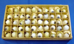
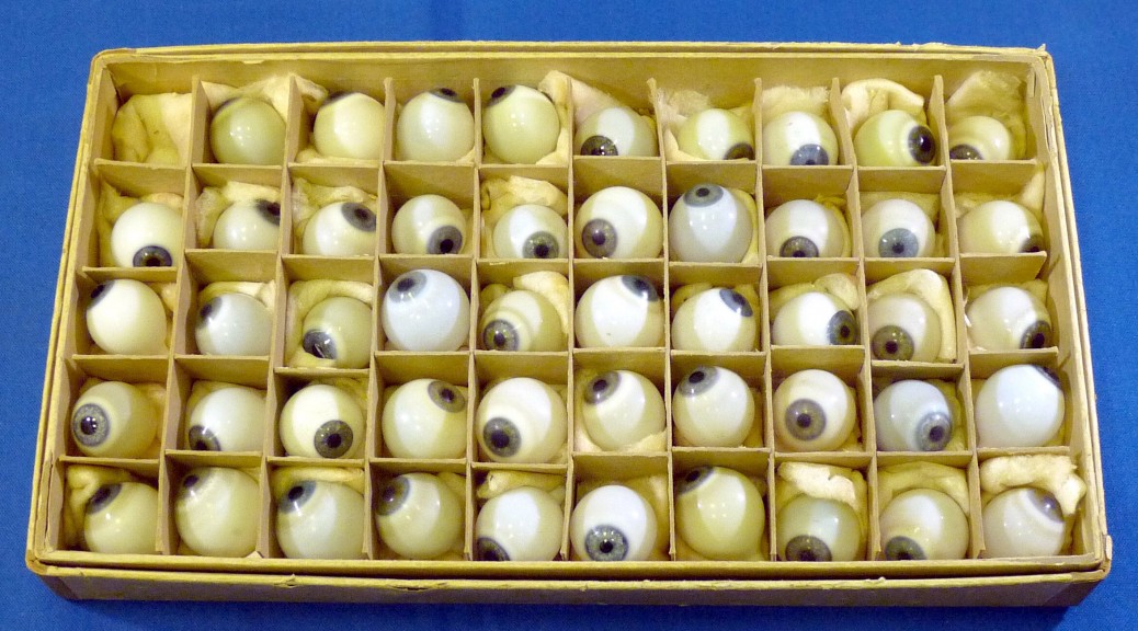
glass eyeballs

Glass eyeballs

Artifacts range in date from the 17th century to today, with many aspects of medicine highlighted in the collection: surgery, gynecology, pediatrics, and more. That “more” category includes items like our prosthetic glass eyeballs, the irises of which are all blue. These hand-blown glass items were intended to serve as prosthetic eyes. In a box reminiscent of a Whitman’s sampler, these eyes were made in a variety of sizes with differing tints of yellow and white. This box would have been used and carried by a traveling salesman of sorts – a person who visited doctors’ offices and sold them to patients in need.

A remarkable collection guide is now available to document the range of historical medical artifacts we have in our holdings.
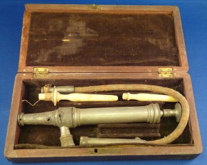
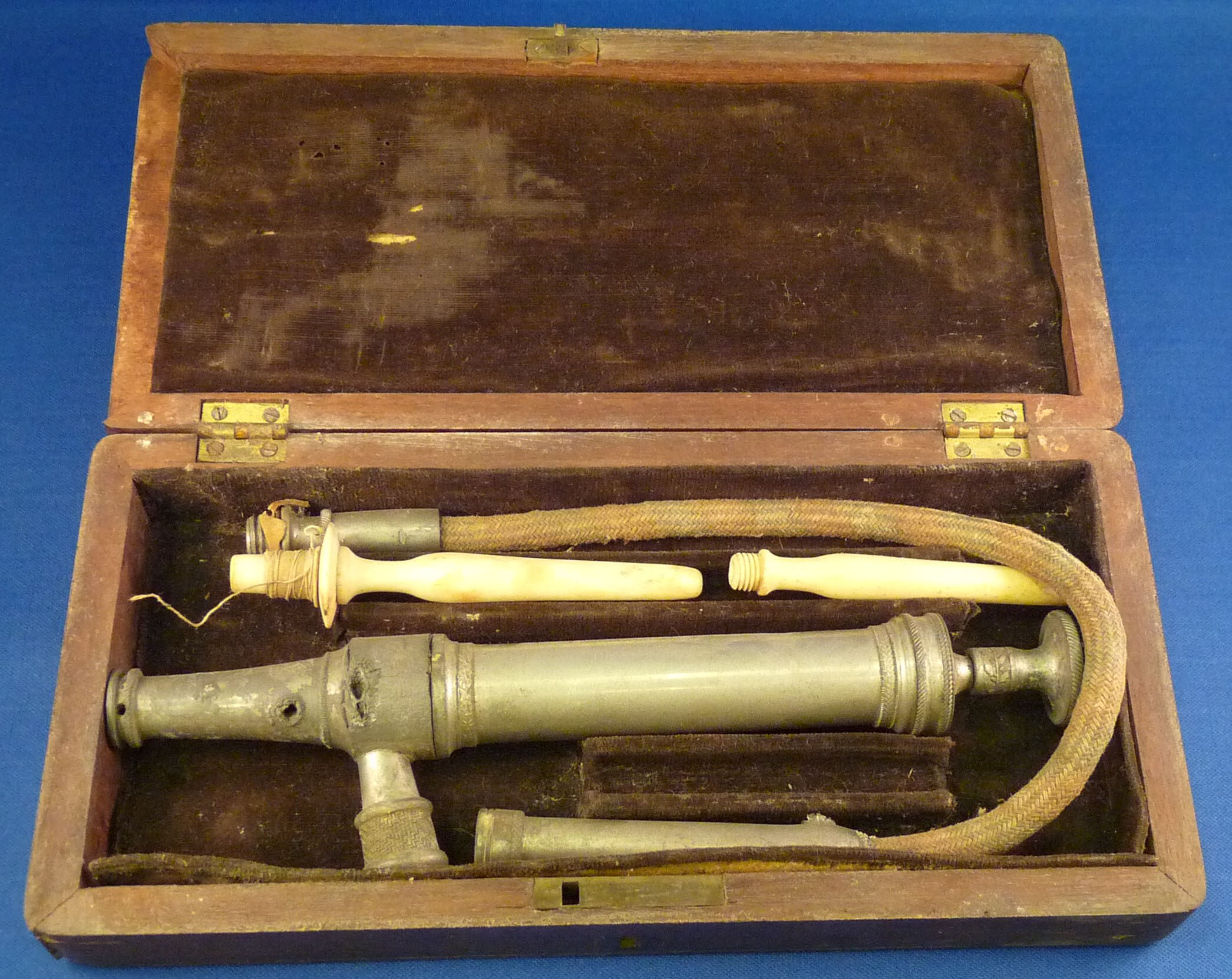
enema syringe

Enema syringe

I’m often asked by students and researchers what my favorite item is. It really changes on a weekly basis. Currently, I’m intrigued by our range of microscopes. They are quite beautiful, span several centuries, and some include ivory specimen slides – with specimens!

microscope

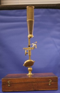
Box mount microscope

 scope2

Screw barrel microscope

Some of my other favorites? Female pills and dental keys. Why were these female pills created and what are they? When I ask students, they often answer that it’s really just Midol.
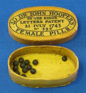
female pills

Female pills

The dental keys really illustrate how medical technology has evolved in tooth extraction. The illustration below shows how a dental key would be used. This image is from the monumental work by J. M. Bourgery, Atlas of human anatomy and surgery, that includes a translation of his work and colored facsimiles. Of note, we also have the nine volume original work of Bourgery.
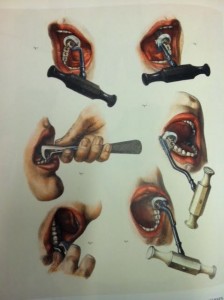
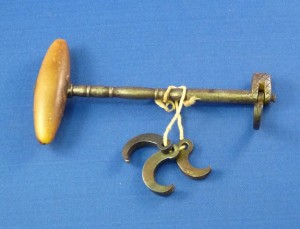
dental chart

key

Dental key

A tremendous amount of thanks go out to so many people: our donors who have generously given such wonderful materials; previous staff of the History of Medicine Collections who researched these items, provided descriptions, and took photos that we can now share; and current staff from the Rubenstein Library’s technical services department and DUL digital projects – all who have made this guide possible.

What will your favorite item be?

Post by Rachel Ingold, Curator for the History of Medicine Collections in the Rubenstein Library.
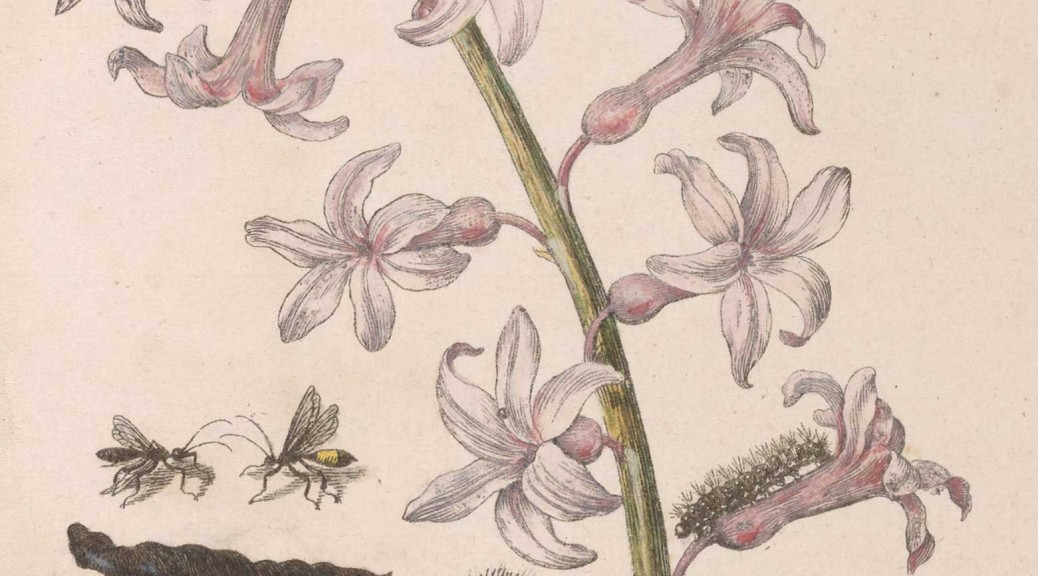