Post contributed by Sarah Bernstein, Josiah Charles Trent History of Medicine Intern.
As someone who studies unorthodox and fringe medicine, I was incredibly pleased to find the large arrangement of unorthodox, fringe, strange, and frankly “quack” medicine within the Rubenstein Library. While the rich History of Medicine Collections includes classics of Western medicine like a first edition of Andreas Vesalius’ De Humani Corporis Fabrica, a memento mori in carved ivory, and various microscopes (on permanent display in the Trent Room), I am glad to share that there are also patent medicine bottles, advertisements, and numerous writings and publications on alternative and unorthodox medicine. George Starr White’s My Little Library of Health is one such series of advice from a so-called “quack,” or an illegitimate and opportunistic, doctor.
The 1928 “little library” by White is a series of 28 books whose length ranges from 20–48 pages. While small, I would say that calling them “thumb-nail” editions is a little misleading; the books measure at 4.5 inches in height and near 3.5 inches across (3 ⁷⁄₁₆ to be exact) is far from what is considered a miniature book or thumbnail sized. The advertisement at the back for each book boasted that each book contained illustrations, sometimes in color, and provided White’s sound advice on “health building by natural living.” Each book could be purchased for 25 cents (now somewhere near $4.50) or, for 5 dollars prepaid (around $90 for us today), one could score for the entire set.
White was a proponent of chromotherapy, light therapy, and heat therapy. In My Little Library of Health he informed his readers about his research and strong belief in the healing properties of Ultra-Red Rays. Although White’s belief in chromotherapy began by viewing sunlight through oak leaves, based on his account in volume 27, his tests had revealed to him that artificial lights from electric lamps still produced healing effects. In fact, some electric lamps worked better than others. Why? Ultra-Red Rays, that White describes as “the ‘thermal’ Rays upon which all life depends,” more commonly known as infrared light. Based on these beliefs, White developed the “Filteray Pad,” a heat pad which generated Ultra-Red Rays and was meant to be applied to the affected area. The price for this cure-all device? A cool $35 (~$620-30 in 2024).
Figure of the Filteray Pad in Volume 28, page 14, of My Little Library of Health (1928).
White would go on to develop other light-based therapies and medical systems. In 1929, White was unflatteringly covered in the “Bureau of Investigation” section of The Journal of the American Medical Association (volume 92, number 15) for his dubious claim of medical schooling and his career in patent medicines. The article lambasted White and all of his medicines and cures. Along with the “Filteray Pad” there was “Valens Essential Oil Tablets” (sold during the 1918 Flu Epidemic for “Gripping the Flu out of Influenza”) and his methods of “Bio-Dynamic-Chromatic (B-D-C) Diagnosis” and “Ritho-Chrome Therapy” (light-based diagnosis and cure using multiple colored rays that were similar to other forms of chromotherapy; the “Electronic Reactions of Abrams” by Albert Abrams and Dinshah Ghadiali’s “Spectro-Chrome” device respectively).
The Bureau of Investigation (formerly the Propaganda for Reform Department) was created as an outgrowth from the Council on Chemistry and Pharmacy to specifically investigate, disprove, and inform the public about fraudulent nostrums and patent medicine. The effort was headed by Dr. Arthur J. Cramp, a passionate doctor who was highly critical of nostrums, patent medicines, and the lax regulations which enabled proprietors to label and advertise their products as legitimate medicines.
George Starr White was just one of many quacks that Dr. Cramp and The Journal of the American Medical Association investigated and denounced, and who are represented in the Rubenstein Library’s collections. While I would not advise anyone to turn to White for medical advice today, I would encourage people to think about illegitimate medical professionals like White—and the world that they operated in—in contrast to medicine and the medical system today. These quacks from the past can provide insight into how medicine is legitimized, the rise of the medical profession, and continuous efforts throughout history to seek and provide unorthodox care.
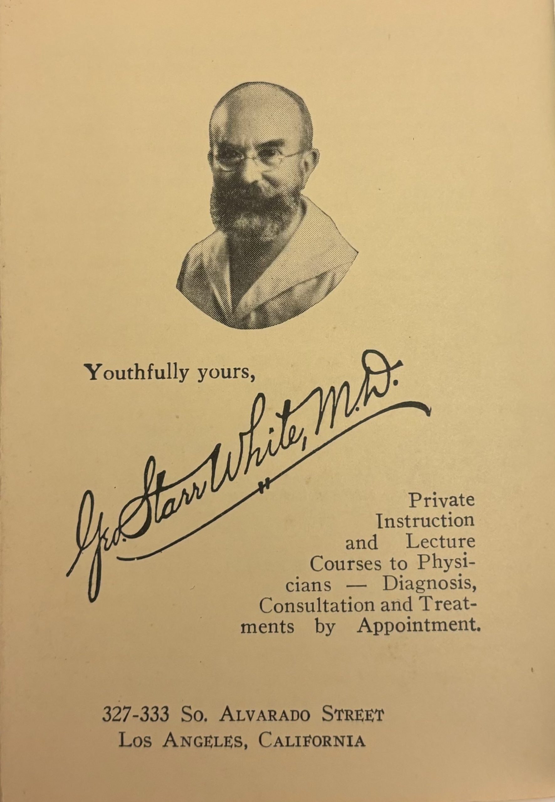
Page with a portrait of George Starr White signed “Youthfully yours” at the end of each My Little Library of Health (1928) book.
Date: Tuesday, January 23, 2024 Time: 5:30 p.m. Location: Rubenstein Library Room 153, Holsti-Anderson Family Assembly Room, Duke University (the event will be recorded) Contact: Rachel Ingold (rachel.ingold@duke.edu or 919-684-8549)
Dr. Kylie Smith
Please join us on Tuesday, January 23, for our next Trent History of Medicine Lecture Series event. Kylie Smith, Ph.D., will present “Jim Crow in the Asylum: Psychiatry and Civil Rights in the American South.”
The Civil Rights movement of the 1950’s and 60’s sought to end racial segregation in all U.S. public institutions, including hospitals. Psychiatric hospitals became political battlegrounds over segregation and patients’ rights, setting the scene for disparities that continue today.
“Jim Crow in the Asylum” explores the process of desegregation and deinstitutionalization in state psychiatric hospitals in Georgia, Alabama and Mississippi. It draws on original records, court cases, and personal testimony to expose the racist ideas that underpinned the treatment of African Americans with mental illness and saw psychiatric hospitals used as dumping grounds for some of the South’s most vulnerable people.
Kylie Smith is Associate Professor and Director of the Center for Healthcare History and Policy in the Nell Hodgson Woodruff School of Nursing, and Associate Faculty in the History Department, at Emory University in Atlanta, Georgia. She earned her PhD in the history of psychiatry in Australia, and is the author of the award winning book Talking Therapy: Knowledge and Power in American Psychiatric Nursing published by Rutgers University Press in 2020. Her new book entitled Jim Crow in the Asylum: Psychiatry and Civil Rights in the American South will be published by UNC Press early in 2025 and is supported by the G13 Grant from the US National Library of Medicine.
This event is sponsored by the History of Medicine Collections in the Rubenstein Rare Book & Manuscript Library and the Trent Center for Bioethics, Humanities & History of Medicine. The Trent Center is holding another talk by Professor Smith on Wednesday, January 24, at noon
Post contributed by Roger Pena, Research Services Librarian
During the Fall 2023 semester, the History of Medicine Collections of the Rubenstein Library welcomed close to 50 first-year graduate students from the Occupational Therapy program at Duke’s School of Medicine. Duke Health and Hospitals have provided occupational therapy services since the 1940s and 2021 marked the inaugural year of the Occupational Therapy Doctorate program.
Occupational Therapists (OTs) are trained in the social, emotional, and physical effects of an illness, injury or disability and help support the development of daily life (occupational) skills to help patients live independently and perform everyday tasks more easily and with less pain. For example, one (of many) functional areas OTs address is handwriting, where providers support skills through physical exercises, self reflection, organizational goals and confidence building.
Although it may be considered a “new” field, with the establishment of the National Society for Promotion of Occupational Therapy (now known as the American Occupational Therapy Association, or AOTA) in 1917, many principles and treatments of occupational therapy can be seen throughout medicine prior to the 20th century in areas such as psychiatry, hygiene, physical therapy and rehabilitation. What may surprise some is the fact that some early interventions of occupational therapy took from the arts and crafts movement of the early 20th Century as well as weaving, gardening and the art of bookbinding. By 1918, occupational therapy schools were established in Boston, Milwaukee, St. Louis and Philadelphia to train reconstruction aides (as OTs were known at the time) in evidence-based practices and treatments to help soldiers returning from World War I. Soon, however, occupational therapy would grow to reach a wider range of patients and those in need of more holistic interventions.
The visit to the Rubenstein Library served as an opportunity for these future OTs to interact with the History of Medicine Collections and Duke Medical Archives artifacts, manuscripts and rare books related to the history of their field and related branches of medicine.
Materials included:
Massage roller and devices to help feed patients with physical limitations
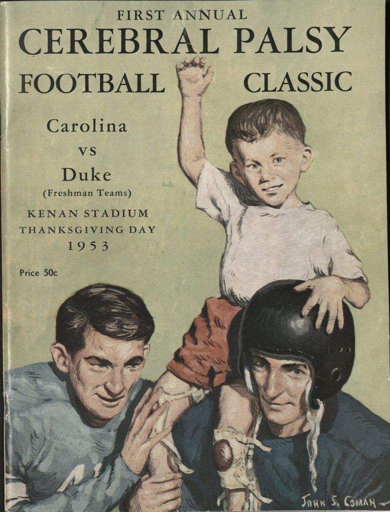
Duke football programs from the 1950s commemorating annual match between Duke and UNC to raise money for the NC Cerebral Palsy Hospital in Durham and raise awareness
Medical illustrations and student journals from OTs
Cover of Duke Football Program, 1953
Duke Football Program, 1953
A Psychiatrist’s Outlet
While curating Rubenstein Library materials for this session, one title of particular (and peculiar) interest was “A Psychiatrist’s Anthology” by Dr. Louis Karnosh, published in 1932 (2nd edition) by the Occupational Therapy Press. This small publisher was part of the formally named Neuro-psychiatry Department at City Hospital in Cleveland, Ohio. Dr Karnosh, a psychiatrist, was the head of the above-mentioned department and also served as a professor of pathology and dentistry at Case Western Reserve University. “A Psychiatrist’s Anthology” – a collection of poems and stories – is inspired by Karnosh’s patients and looks at six different psychiatric conditions suffered by those he is treating – delirium tremens, general paresis, melancholia, schizophrenia, paranoia, and senile dementia.
The anthology also serves as an ode to his love of poetry and book printing. The first part explains Karnosh’s reasoning for creating an anthology of poems and stories while also describing his desire to publish a book in the tradition of Old World book binding and printing. “As a specimen of bookcraft, this is but an amateur’s feeble emulation of master bookmakers of yesterday … The pen and ink sketches, the type composition and printing are done by the author.” The uncut pages, woodcut illustrations, the typography of the movable type, and the limited numbered copy give the air of books printed centuries before.
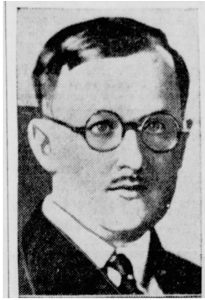
Dr. Karnosh in 1940
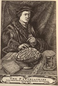
“Self-portrait of a psychiatrist”
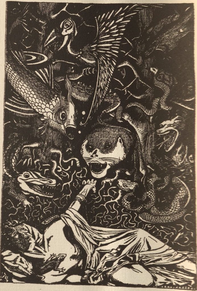
Every part of this anthology has symbolism and meaning to represent the six conditions Karnosh delves into through story and rhyme. The preface is written in poetic verse and explains Karnosh’s thoughts and ideas on what it means to be a psychiatrist while the introduction gives Karnosh the opportunity to speak to his readers about empathy for his patients – to not convolute mood with madness. ” I must keep to the road… Luring sirens… are calling and are singing phantastic farrago of popular psychologies. I must retain an objective calm…. I must first be an able dissector before I can synthesize…. Above all I must not treat diseased effectively by interjecting my own into the problem at hand. There must be no clash of feeling with feeling.” Each of the six conditions includes an original hand-drawn illustration, poem, and patient anecdote by Karnosh as well as a short encyclopedia entry about a mythical or literary figure to help the reader better connect to the condition(and patient) described. The folly of Prometheus to describe delirium tremens (alcohol withdrawal); Don Quixote to describe the condition of paranoia.
The (Poet) Doctor is In
Newspaper article reporting Karnosh attack
Physicians as poets and writers is a tradition that dates back centuries, from John Keats, William Llyod Carlos and Oliver Wendell Holmes Sr. to the contemporary Nawal El Saadawi, Irène Mathieu and Rafael Campos. Physicians have long seen the therapeutic value of writing and reading poetry for both patients and themselves. At Philadelphia Hospital, founder Benjamin Franklin and Dr. Benjamin Rush were proponents of patients using writing as therapy while Walt Whitman was known to visit field hospitals and read poetry to wounded soldiers during the Civil War.
A look into the life of Dr Karnosh revealed an interesting and almost haunting past. Aside from his responsibilities as the head of the Neuropsychiatry Department at Cleveland Hospital and medical school professor, Dr Karnosh served as an expert witness in the Cleveland area murder cases[1] for both prosecutors and defense teams, having the ultimate responsibility of determining the mental capacity of the accused to stand trial. In 1940, Dr Karnosh was shot[2] on the front door of his home by a former patient who had accused him of writing a negative evaluation that resulted in the loss of the patient’s pension. Karnosh survived the attack.
Writing and poetry seemed to have had a therapeutic value for Dr Karnosh and may have served as an outlet for all the stress and pressure that came with his responsibilities. He was known to give lectures on various topics across the Ohio area and contributed advice columns[3] in local papers. A look at the catalog record at Case Western Reserve University’s Library, shows several books attributed to Karnosh as the main author, co-author or medical illustrator, including a textbook on psychiatry for nurses that had eight different editions.
As for the link between physician and poetry, Dr. Rafael Campos, physician, poet and Director of Literature and Writing Programs of the Arts and Humanities Initiative at Harvard Medical School explains, “Poetry does a better job in teaching because it is about embracing the human aspect of suffering.” He goes on to say, “It’s our own humanity. That can be really healing for patients.”[4]
A New Approach
In explaining his thoughts for writing a “Psychiatrists Anthology,” Karnosh mentions that it is a “product of avocational moments of a psychiatrist who spends time with his patients in occupational therapy.” By the time the first edition was printed in 1931, occupational therapy had been an officially recognized medical discipline for close to fifteen years but “didn’t fit neatly into the medical model.” OT took a more holistic approach to therapy with aspects of nursing care, physical therapy, social work, psychiatry, health advocacy, and orthopedics seen in its treatments and interventions. Were poetry and art how Karnosh connected with his patients? Were these occupational skills part of his own mental well-being?
At a time when those with mental illness, physical disabilities and depression were seen as ills of society, occupational therapy and new OTs seemed to bring a breath of freshness to medicine with a focus on developing vocational skills as well as supporting the mental well-being of patients.
Today, there are approximately 500 accredited Occupational Therapy programs (with nearly 200 more vying for accreditation status) and over 180,000 occupational therapists and OT assistants practicing in schools, hospitals and outpatient clinics across the United States. As of December 12, 2023, Duke University’s Occupational Therapy Doctoral Program was granted accreditation from the Accreditation Council of Occupational Therapy Education (ACOTE).
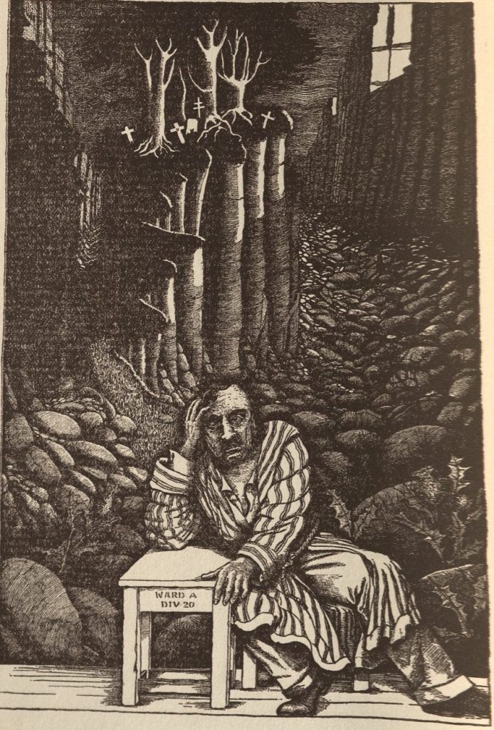
Illustration by Karnosh from “A Psychiatrist’s Anthology”
Illustration by Karnosh from “A Psychiatrist’s Anthology”
Illustration by Karnosh from “A Psychiatrist’s Anthology”
Footnotes
[1] The Evening Review. East Liverpool, Ohio. Tuesday, January 26, 1932
[2] The Daily Sentinel-Tribune.Bowling Green, Ohio. Wednesday, July 03, 1940
[3] The Coshocton Tribune. Coshocton, Ohio. Thursday, October 12, 1939
Harry H. Harkins T’73 Travel Grants for Lesbian, Gay, Bisexual, and Transgender History
History of Medicine Collections
Human Rights Archive
John Hope Franklin Research Center for African and African American History and Culture
John W. Hartman Center for Sales, Advertising & Marketing History
Sallie Bingham Center for Women’s History and Culture (Mary Lily Research Grants)
Anyone whose research would be supported by sources from the Rubenstein Library’s research centers is eligible to apply. We encourage applications from students at any level of education; faculty and teachers; visual and performing artists; writers; filmmakers; public historians; and independent researchers. For assistance determining the eligibility of your project, please contact AskRL@duke.edu with the subject line “Travel Grants.”
Eligibility
Applicants must reside beyond a 100-mile radius of Durham, N.C., and may not be current Duke students or employees.
Information Session
An online information session will be held Thursday, January 11, 2024, 2-3 pm EST. This program will review application requirements, offer tips for creating a successful application, and include an opportunity for attendees to ask questions. This program will be recorded and posted online afterwards. Register for the session here.
Timeline
The deadline for applications will be Thursday, February 29, 2024, at 6:00 pm EST.
Decisions will be announced by the end of April 2024 for travel during May 2024-June 2025. Awards are paid as reimbursement after completion of the research visit(s).
14 June 1850 resolution of the Joint Committee of the Library of Congress, Box 15, Wilkes Papers
Upon successfully passing the motion at their meeting in June 1850, the Joint Committee of the Library of Congress resolved to compel Charles Wilkes to “notify Mr. Pickering that the Committee think he was not authorized to devote his time” as a member of the United States Exploring Expedition between 1838 and 1842 to jotting notes for his book The Races of Man.[1] Nevertheless, Pickering published The Races of Man as the ninth volume of the multi-volume Narrative of the United States Exploring Expedition in 1848, six years after returning from their voyage under the command of Lieutenant Wilkes. The committee’s resolution to Wilkes and Pickering is among the Wilkes Papers held by the David M. Rubenstein Manuscript and Rare Book Library, which generously funded my research at the library in the summer of 2023.
During their time in the Pacific Ocean––including stopovers in the Tuamotu Archipelago, Tuvalu, Tahiti, Samoa, Fiji, Hawaii, and the Philippines––Pickering resolved to produce a classificatory schema of “all eleven races of man.”[2] At the start, he found “difficulty arose, in fixing in the mind, while passing from place to place, the relative shades of complexion” of the people the Exploring Expedition, or Ex. Ex., encountered during their voyage.
Fijian skin, for instance, upset English-speaker’s reliance on vision to discern race in the early nineteenth century. In May, 1840, Pickering looked through a spyglass from the deck of the Vincennes, the squadron’s flagship, toward a cluster of people gathered on the shore of Levuka, a town on the eastern coast of Ovalu, to obtain “evidence of the lightness of the Feejeean complexion.” Ovalu is one of the more than three hundred volcanic islands that make up the Fiji archipelago in the South Pacific.
At first, Pickering incorrectly hypothesized the group contained a mixture of “Malayan”, “Polynesian”, and “Negro” peoples rather than Fijians. Seeing people from afar thus proved to be inadequate for the purposes of collecting scientific facts concerning skin color in the Pacific Rim. Pickering improvised by terming them “purple men” on closer inspection. Ocularity and visibility, then, proved to be incomplete methods for knowing race.[3] So, Pickering concluded, his racial scientific program required collecting “more obvious distinctive characters” to serve as an evidentiary basis for his racial taxonomy. Some of these characters included notes on Papuan skin as “harsh to the touch, and the hair crisped or frizzed”, hearing Pa‘umotus “making a kind of purring noise”, and wincing at “the strong ill odour” of Fijians that “make them thoroughly disgusting to persons newly arrived.”[4]
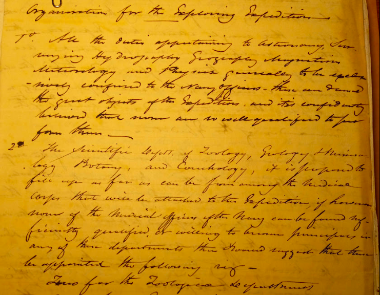
“Organization for the Exploring Expedition”, Box 3, Folder 1, Wilkes Papers
Pickering’s inability to fully rely on vision matters for historians of science and the senses. Relying on prior analyses of race as a phenomenological apparatus, in particular the scholarship of philosophers including Sachi Sekimoto and Christopher Brown, I am investigating how the Ex. Ex. produced scientific ideas about race via the sensorium. What is at stake here is the place of vision and visibility in histories of science in the Enlightenment as hallmarks of modern scientific epistemology. Forms of visualization equipped what Lorraine Daston and Peter Galison term the disciplinary eye that lay at the ethico-epistemic foundations of contemporary science.[5] Yet, scientists like Pickering used hearing and ideas about noise, smell and notions of cleanliness, and mores around touch and taste, to articulate race as a scientific fact through the itinerary of the Ex. Ex. Put simply, ocularcentrism was too brittle an epistemological basis for the Ex. Ex. to taxonomize the various groups they “discovered” through their transpacific itinerary. Rather, the Ex Ex used olfactory disgust, sonic boundaries, and norms surrounding touch and gustation to classify Pacific Islanders as racialized others through the body and the senses.
Before the Ex. Ex. departed from Hampton Roads in 1838, Wilkes argued that the operation would prove to be “useful to the Navy, honorable to this Country, and highly advantageous to the Commercial interest of the Country” and to “Science generally.”[6] In his “Organization for the Exploring Expedition”, Wilkes did not propose sending a race scientist like Charles Pickering––who joined the Ex. Ex. as the scientific corps’s zoologist––along with the other “Scientifics” like the geologist James Dwight Dana, the botanist William Rich, or the artists Alfred Thomas Agate and Joseph Drayton.[7] The Wilkes Papers at the Rubenstein contain material on these figures, as well as the John Torrey Papers, which pertain to the Ex. Ex. Torrey––a botanist who did not travel with Wilkes––later classified the plant collections made by the scientific corps and prepared specimen catalogues as an affiliate of the Smithsonian Institution, and his papers contain letters with people associated with the SI like Spencer F. Barid, Joseph Henry, and Louis Agassiz. Torrey’s correspondence also contains letters from the phrenologist Johann Gaspar Spurzheim, and Josiah Nott, a leading race scientist of the antebellum era.
Moving forward, my aim is to produce a phenomenological account of the Ex. Ex. that provides insight into the formation of the racist ideas that undergirded Indian removal and Manifest Destiny via the senses. Like Sachi Sekimoto––who argues that “race constantly renews its material presence through latching onto our bodily felt, sensorial experiences, making itself feel-able and sensible and therefore ‘natural.’”––I claim that the narratives produced by the scientific corps and the naval personnel of the Ex Ex justified beliefs in American Indian and Polynesian “savagery” in Jacksonian America.[8]
[2] Charles Pickering, The Races of Man: And Their Geographical Distribution (London: H. G. Bohn, 1850) 2nd edition, 2.
[3] Charles Pickering, The Races of Man: And Their Geographical Distribution (United Kingdom: John Chapman, 1849), 146-147.
[4] Pickering, The Races of Man, 3; Wilkes, Narrative of the United States Exploring Expedition, vol.1, 324; Walter Lawry, Friendly and Feejee Islands: A Missionary Visit to Various Stations in the South Seas in the Year MDCCCXLVII, (United Kingdom: C. Gilpin, 1850), 79-80.
[5] Lorraine Daston, and Peter Galison, Objectivity (Princeton: Zone Books, 2007), 48, 148
[6] Wilkes Papers, Box 3, “Organization for the Exploring Expedition”
[7] William Reynolds, Voyage to the Southern Ocean: The Letters of Lieutenant William Reynolds from the U.S. Exploring Expedition, 1838-1842 (United States: Naval Institute Press, 1988), 3.
Post contributed by Rachel Ingold, Curator for the History of Medicine Collections.
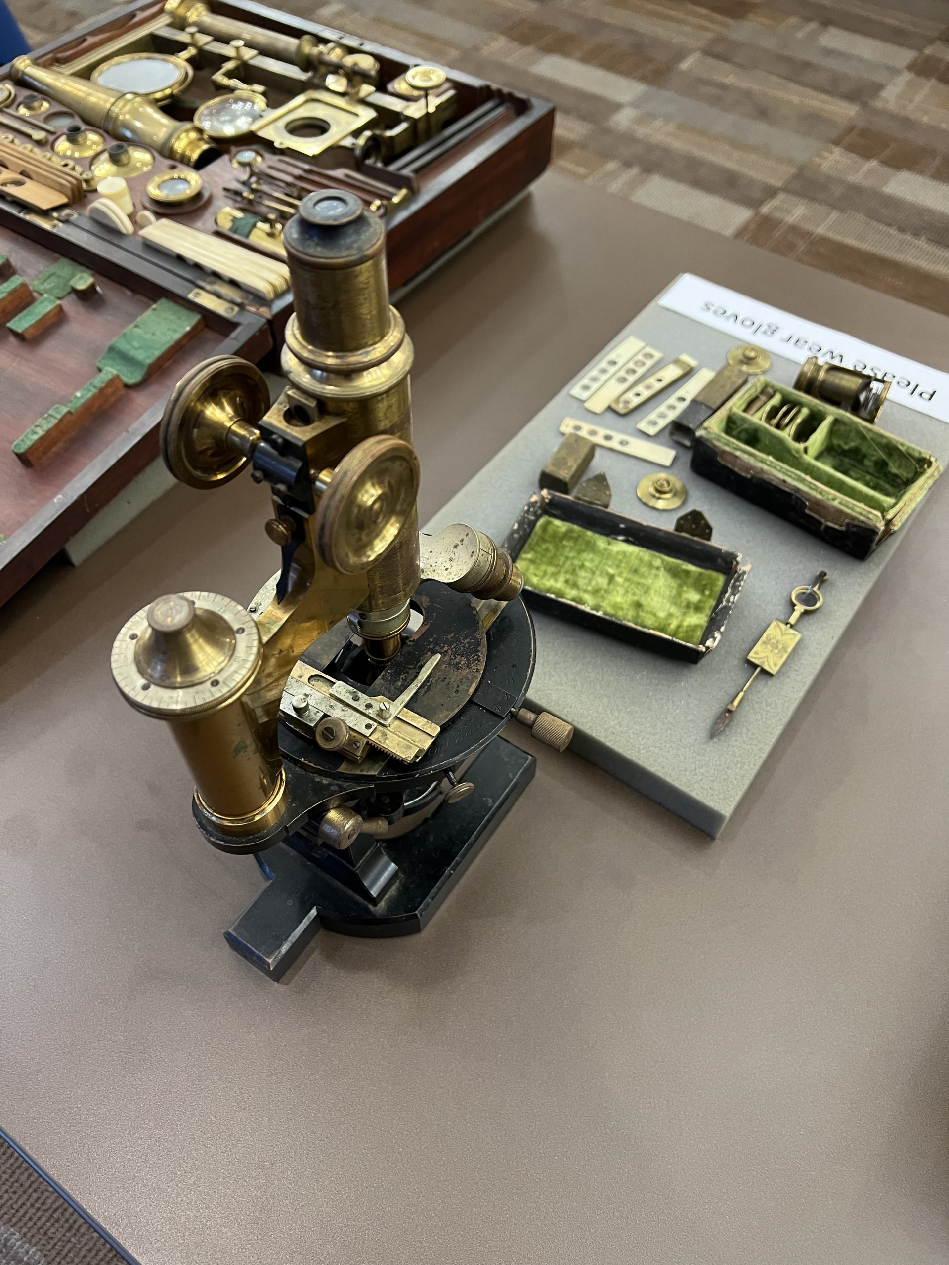
In September, the Rubenstein Library partnered with colleagues in the Natural and Engineering Sciences (NSE) for an open house event. While our Engineering Exposition targeted students, faculty, and staff from Duke’s Pratt School of Engineering, all were welcome to attend.
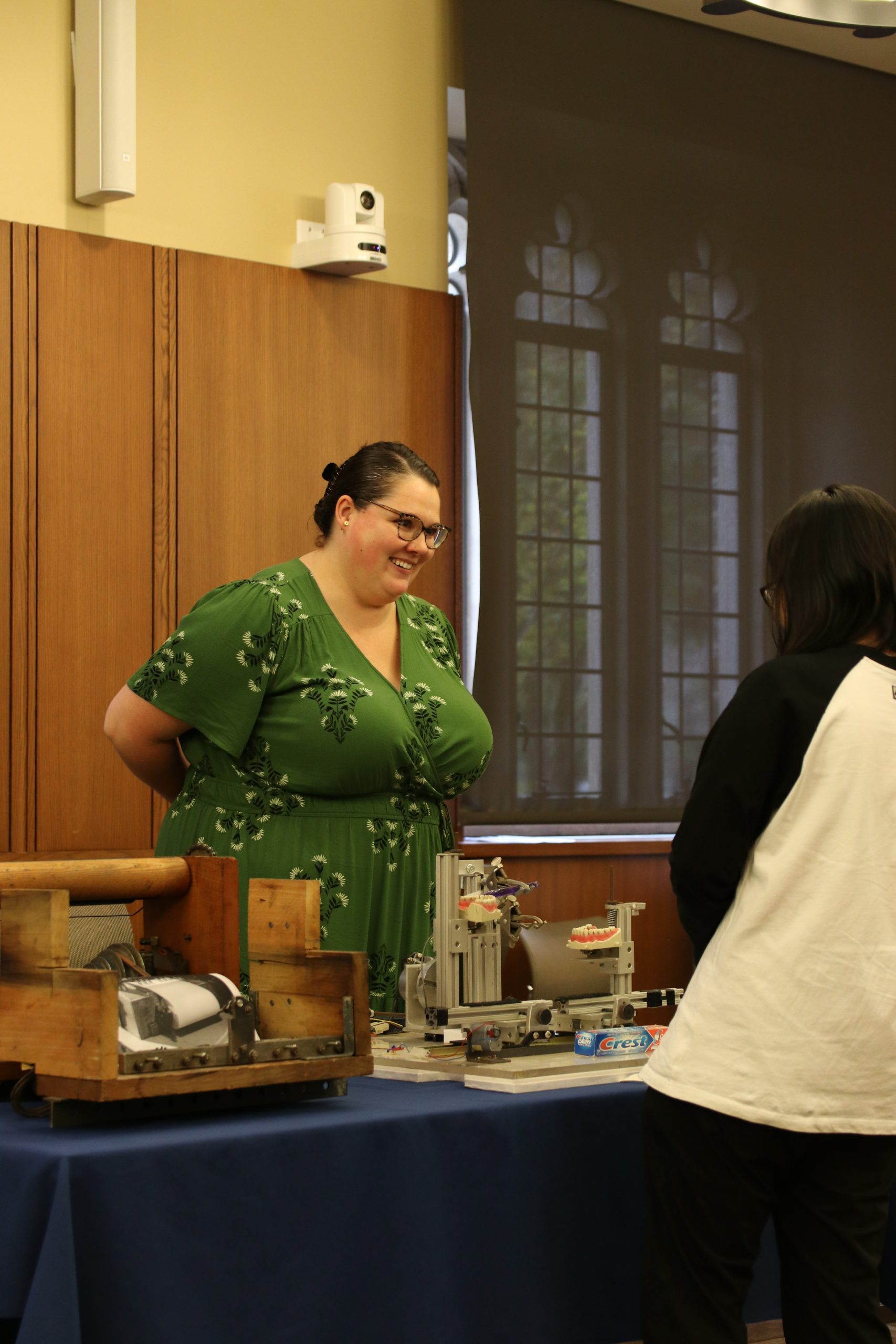
Faculty from the Engineering School examine works on engineering from the 16th and 17th centuries! Photo by Deric Hardy.Robin Klaus, graduate Intern in the Hartman Center for Sales, Advertising, and Marketing History talks with a student about the toothpaste testing device found in the Consumer Reports archive. Photo by Janelle Hutchinson.
Items from a variety of collecting areas within the Rubenstein Library were available for visitors to examine and handle. Some highlights included
And much more! So much more! Including this video from the Consumer Reports lab.
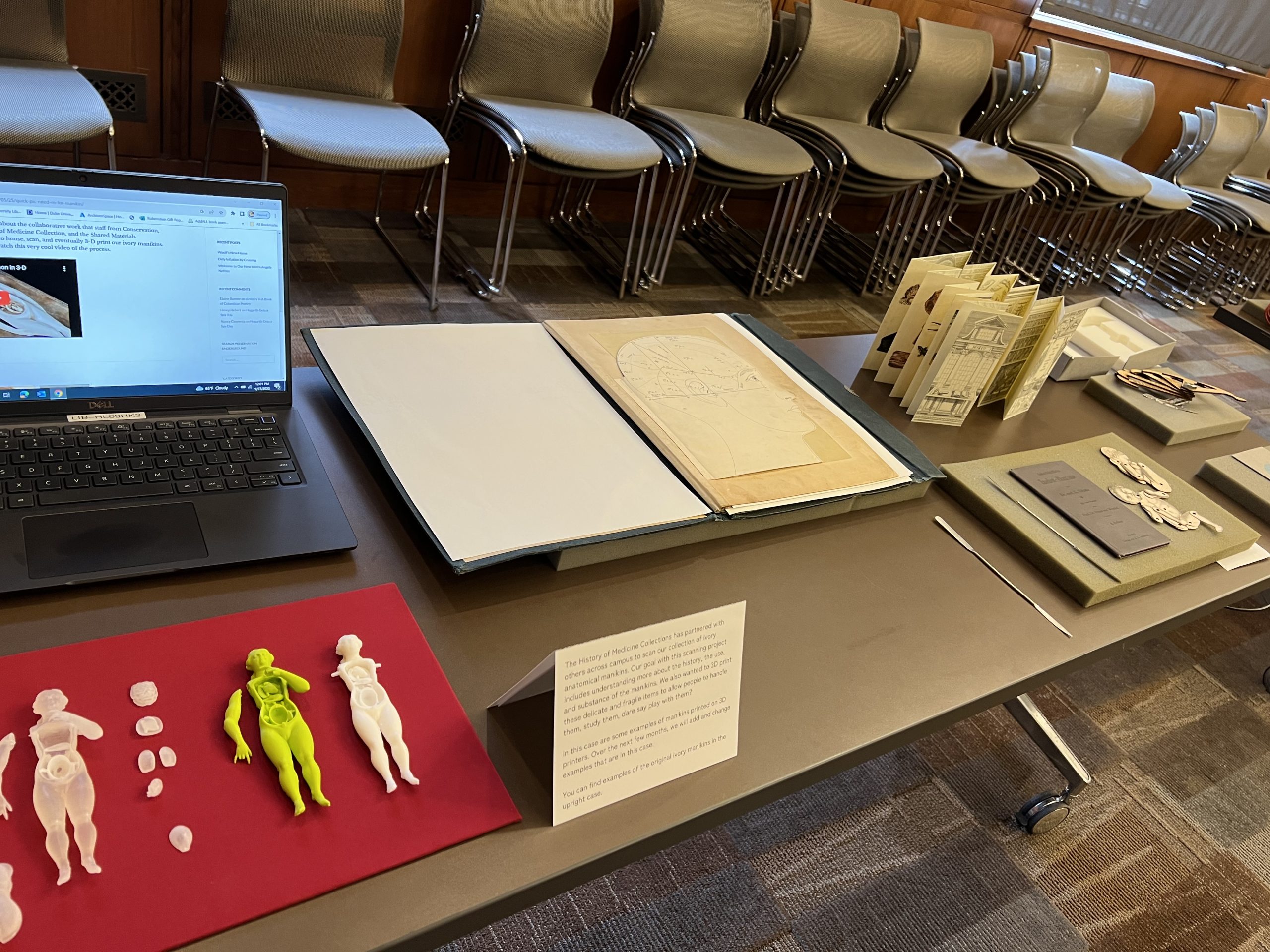
Examples of 18th and 19th century microscopes that visitors were encouraged to handle and try out! Photo by Deric Hardy.Examples of moveable books from the History of Medicine Collections and samples of 3D printed anatomical manikins made from items in our collection! Photo by Deric Hardy.Pages from an anatomical flap book where the flaps can be lifted, as shown on the right, to reveal detail about the human body. Photo by Janelle Hutchinson.
We look forward to our continued partnerships with colleagues across the Library and campus. Let us know what you might like to see at our next Engineering Exposition!
The History of Medicine artifacts collection presents such a unique opportunity to work with material sources in the history of medicine. In the same way that there is a difference between viewing manuscripts through photographs and seeing them in person, there is something striking about being able to hold an object that you have only read about in books and pamphlets. In my training as a historian, I have been largely trained and relied on primary sources in the form of written materials. It is precisely because of this that I have been thrilled to be able to view and work with the History of Medicine artifacts collection.
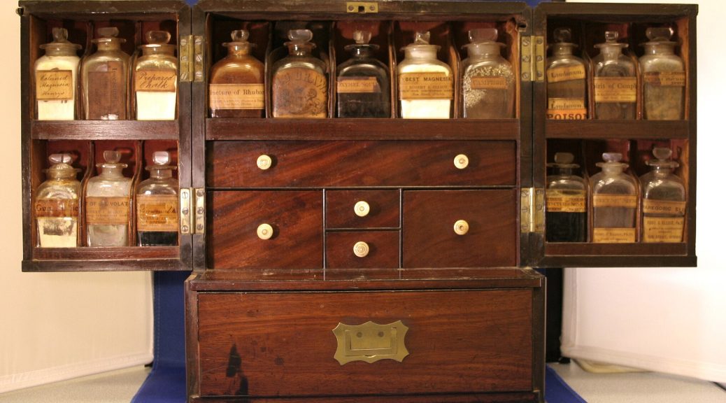
Amongst the many marvelous and unexpected items in the collection, from amputation sets and bone saws to carved ivory manikins and elaborate anatomical flap books, I found myself drawn to the multiple British nineteenth century medicine chests within the collection. These stately century solid wood boxes contained custom glass bottles, fitted to each box’s measurements, with some still filled with powders and liquids. Going through them was nothing short of opening a time capsule and a treasure chest at the same time.
Medicine chests like these can provide a window into the past to understand not only nineteenth century medicine, but global, local, and cultural developments as reflected in the items in these chests and the existence of these chests themselves. There are some medicine chests that are smaller than others, with a variety of cork-stoppered bottles, and were likely meant to be portable and used while traveling. Other medicine chests are heavier and equipped with preparatory tools and medical instruments. These large medicine chests were meant to be stationary, within homes or on ships. In England, both types of medicine chests emerged in the context of newfound social and physical mobility for the Victorian public.
Advertisement from the back of a book within the Rubenstein Library collection, How to Live in Tropical Africa (1912) by John Murray, for a travel medicine chest made of metal.
Regardless of whether they were meant for travel or to be stationary, the existence of these chests speak to the common practice of self-healing, an anticipated absence of a physician, an expected level of medical literacy, and an interest in maintaining one’s own health. These chests are more similar to our contemporary medicine cabinets and in the household, functioned less like a first aid kit or a form of triage support. Rather than immediately, and always, calling upon a doctor, people would often utilize herbal and botanical knowledge to create remedies at home to alleviate and treat their ailments before turning to a physician. And what exactly did people use as medicine?
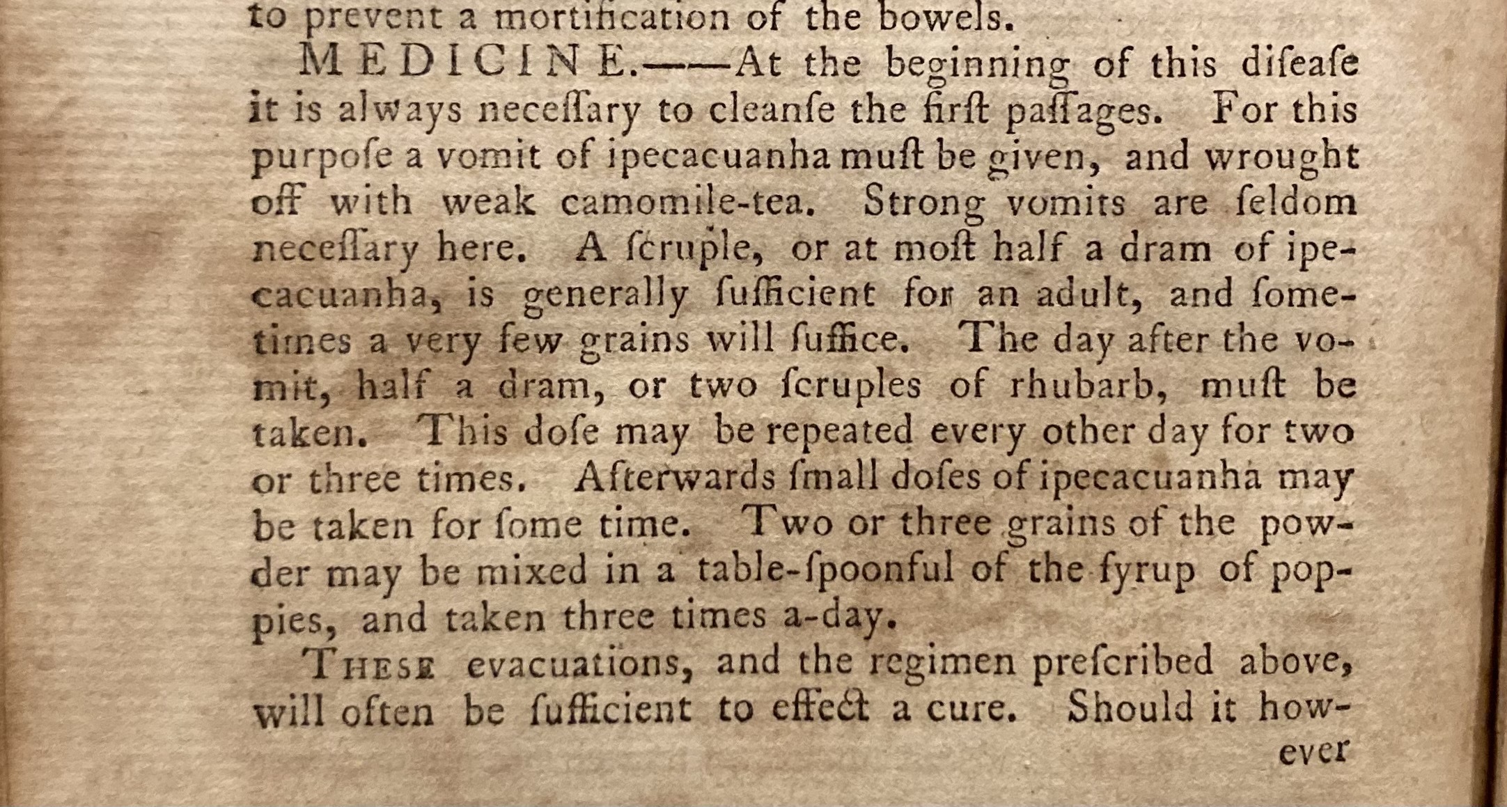
In one “home medicine chest” there are bottles of Ipecacuanha (Carapichea ipecacuanha) in various forms. Ipecacuanha is a slow growing plant native to Central and South America that has a long history in British medicine as to treat dysentery, poisoning, fever, and colds. It was commonly prepared as syrup of ipecac, or simply “ipecac,” which would be used to empty the stomach to combat poisoning. Ipecacuanha was also used in Dover’s Powder, a bottle of which also appears in the same home medicine chest, which was a mixture of powdered ipecacuanha, potassium sulfate, and powdered opium as a pain reliever and to treat fevers and colds by inducing sweating.
Mention of ipecacuanha and rhubarb to treat dysentery in an American second edition of William Buchan’s Domestic Medicine (1774) held in the Rubenstein History of Medicine Collection.
The same home medicine chest also contains multiple instances of rhubarb: tincture of rhubarb, one simply labeled as “Rhubarb,” and the other specified as “Powder of Turkey Rhubarb.” While today rhubarb may conjure thoughts of confectionery sweets and strawberry and rhubarb pie, rhubarb has historically been prized for its medicinal properties and was highly sought after. Rhubarb itself refers to a species of plant, Rheum palmatum, that native to parts of western China and northern Tibet. It was used to aid in cases of indigestion and as a laxative.
Similarly to ipecacuanha, rhubarb and its various preparations can reveal the rich history and practice of herbal and botanical medicine that persisted into the nineteenth century. Despite both of the plants being non-native to Britain, where these chests were created and their clientele were located, ipecacuanha and rhubarb were popular and common treatments utilized throughout the nineteenth century. The prevalence of ipecacuanha and rhubarb not only serves as an indication of the widespread use of purgative medicine during that era but also hints at the emergence and growth of industries, trade networks, and international relationships necessary for the accessibility of these medicinal plants.
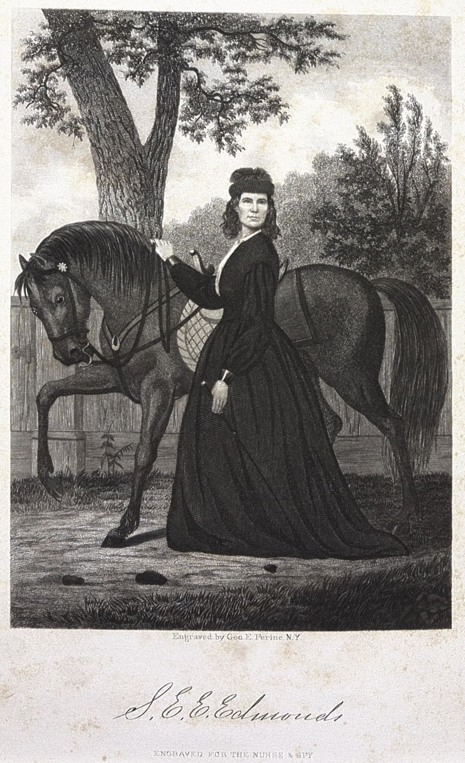
Emma Edmonds, from Nurse and Spy in the Union Army: Comprising the Adventures and Experiences of a Woman in Hospitals, Camps, and Battle-Fields, 1865.
I zipped around the book to get a sense of Emma Edmonds’s time during the Civil War. She was a nurse and when a need arose to infiltrate the Confederate army, Edmonds stepped up. Edmonds went through a process to test her abilities, and a line that stood out to me was regarding her phrenological examination—it showed that she was capable of being a spy.
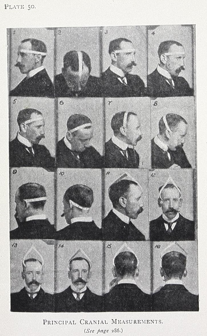
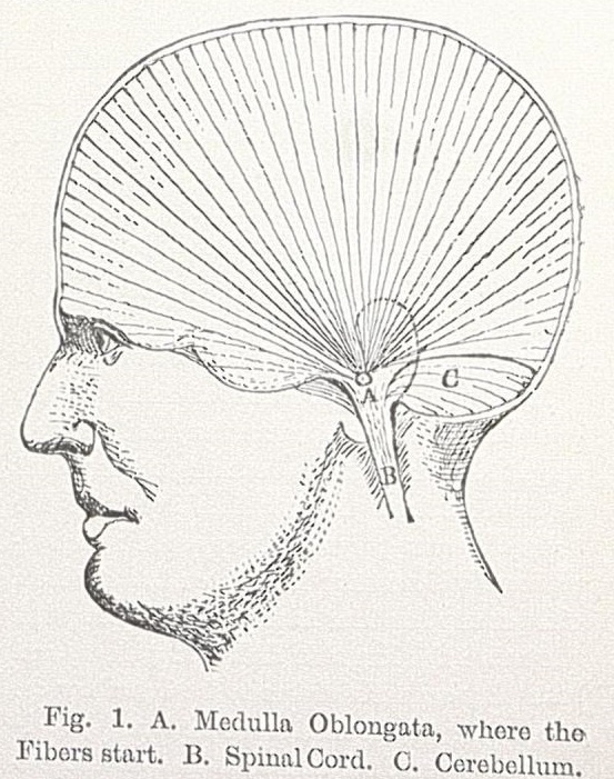
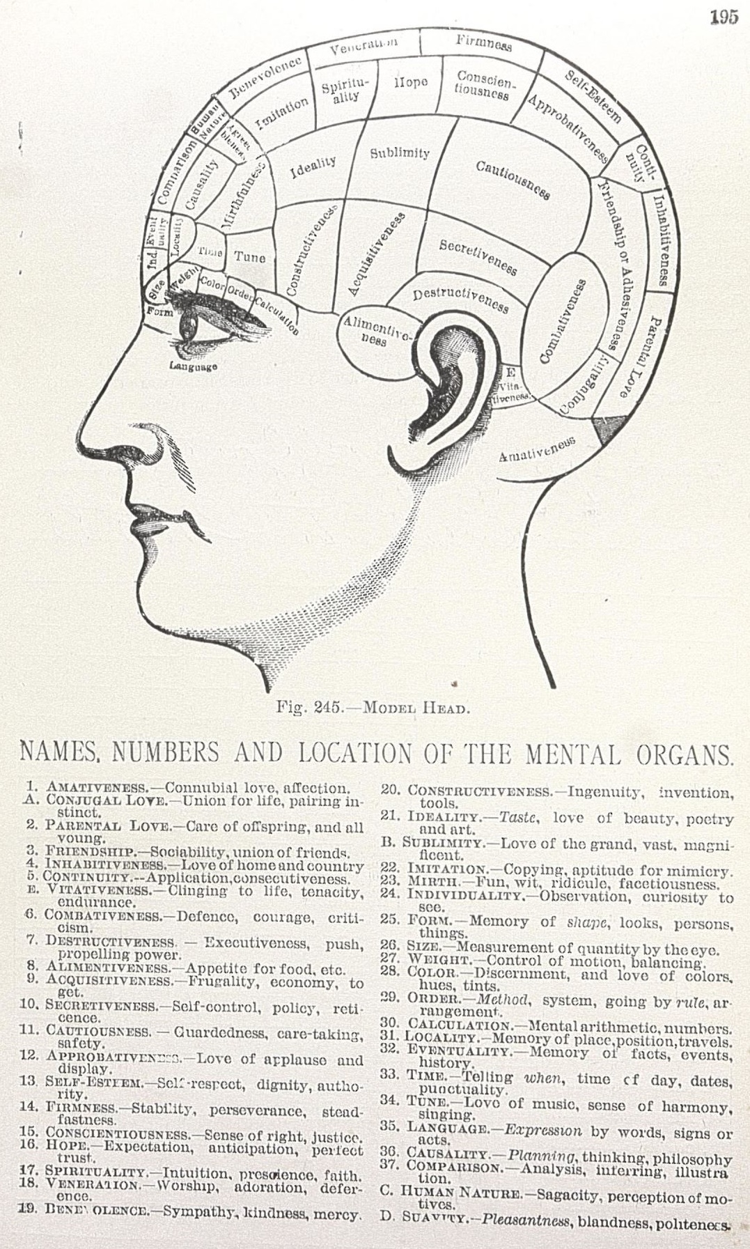
Phrenology has been covered here on the Devil’s Tale before, such as in this excellent post about the phrenology of the Dukes, and the History of Medicine collection includes several phrenological books to enlighten us further. To sum up, phrenology claimed to discern the strengths and weaknesses of a person’s character by measuring the distances from the top of their spinal cord (around the opening of the ear) to the surface of the head, with different characteristics assigned to different parts of the brain/regions of the head. Scientific Phrenology: Being a Practical Mental Science and Guide to Human Character, an Illustrated Textbook by Bernard Hollander, offers a guide on cranial measurements that one should start with their children at six months and go until the age of puberty.
Cranial measuring image from Scientific Phrenology: Being a Practical Mental Science and Guide to Human Character, an Illustrated Textbook, 1902.
People could participate in readings out of their own interest, to check their compatibility with a suitor, to aid in the raising of their children, and phrenologists also played a part in court cases. The pseudoscience’s popularity overlapped with the American Civil War, and apparently also guided in the hiring of spies.
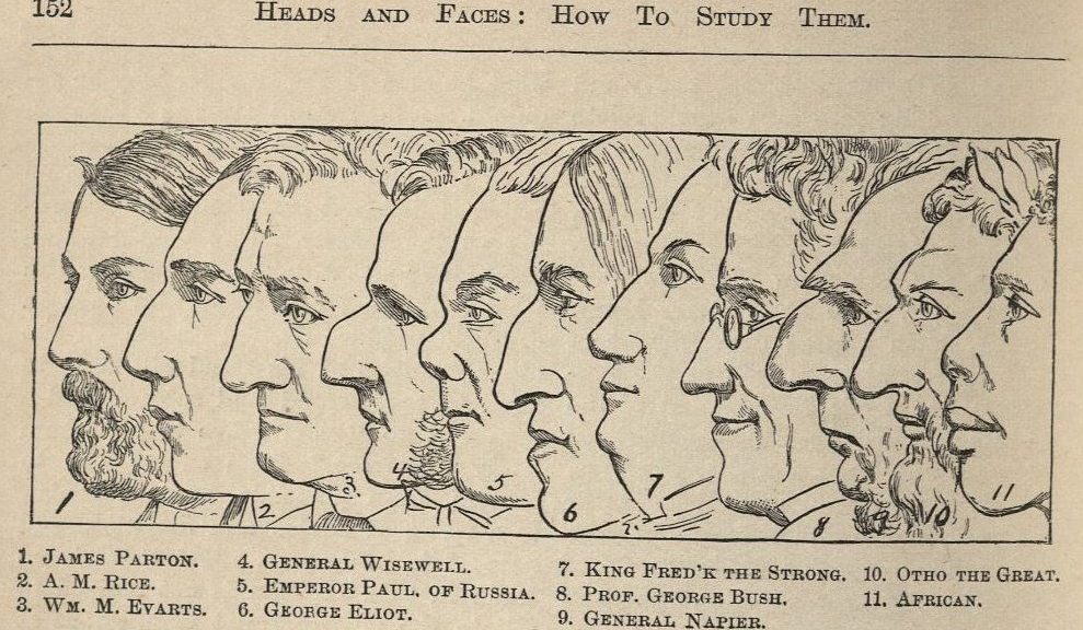
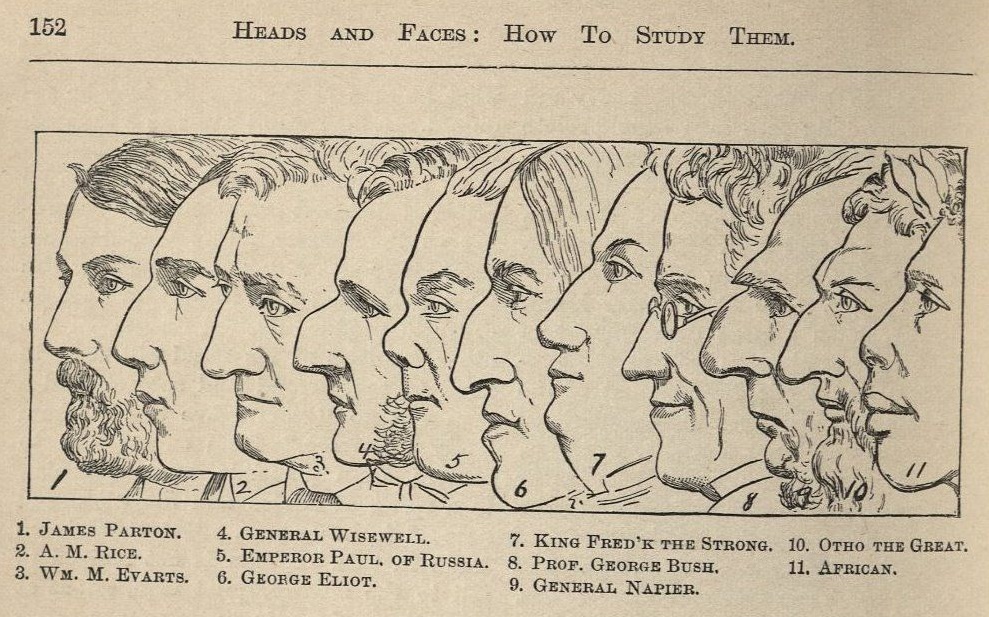
Heads and Faces, and How to Study Them, a Manual of Phrenology and Physiognomy for the People by Nelson Sizer and H.S. Drayton give us a breakdown of the characteristics phrenology covers.
Measuring from the spinal cord to the head from Heads and Faces, and How to Study Them, a Manual of Phrenology and Physiognomy for the People, 1887.
Edmonds mentioned her phrenological exam found her organs of secretiveness and combativeness to be largely developed, then included a vague “etc.”. Regarding secretiveness and combativeness, Sizer and Drayton define it as:
Combativeness. Meets duty bravely, has moral courage, intellectual enterprises, energy of character
Secretiveness. They do not say or do anything in an open, frank manner, but it is by concealment, by artifice, and there is mystery in all they do
Facial profiles from Heads and Faces, and How to Study Them, a Manual of Phrenology and Physiognomy for the People, 1887.
I then decided to make some guesses on what the “etc.” might include. If I were to guess at the qualities Edmonds was strong in (and I don’t mind guessing because this is quack science anyway, though my enthusiasm was tempered by the gross racism found rampant in phrenology and physiognomy), I would guess the following:
Inhabitiveness. Love of home, patriotism;
Self-esteem. Gives confidence in the exercise of courage and judgment;
Firmness. Working with Combativeness, it produces determined bravery;
Imitation. This attribute mostly calls for people to become more refined by imitating others, but it also refers to imitation in common modes of doing and acting;
Individuality. Eager to see all that may be seen and nothing escapes their attention;
Locality. Remembers where things or places are in respect to themselves; they will remember roads and places and directions in a town (here is where I would completely fail as a spy);
Time. Remembers dates and times but also has a sense of time/how long things take; and
Finally, I think Edmonds would have been low on Cautiousness, which can cloud over all manifestations, paralyzing courage, energy, determination, and Hope.
Model head image from Heads and Faces, and How to Study Them, a Manual of Phrenology and Physiognomy for the People, 1887.
Lest we get too excited about lady nurses/spies and their exciting phrenological aspects, Scientific Phrenology reminds us that a woman like Edmonds is an exception, because as Hollander says, the average woman is less intellectual and more emotional than the average man “because of their mammary glands […], their sexual organs being concealed in the pelvis […]”, and various differences in their brains, such as their smaller frontal lobes.
Oh, phrenology, also a friend to misogyny.
This sort of reasoning is, of course, one of the reasons I seek out women in medicine, science, and life. And so we do not end on the sour note of misogyny, you can find meaningful resources on this LibGuide about women and their work in science and medicine.
From the Florence Small Gaynor scrapbook, 1970-1972, manuscript.
Post contributed by Michelle Wolfson, the 2022-2023 Josiah Charles Trent History of Medicine Intern.
Tell us a little bit about yourself.
I am currently studying library science at East Carolina University. I started the program after realizing both of my children would be in school, in-person (the youngest did kindergarten virtually!), and I could get back to work. I enjoyed being a homemaker for nearly a decade; my children are 10 and 8 years old. Instead of returning completely to work, I decided that I did not want my children to one day say about me, “She really wanted to be a librarian but she never did it,” and so I started at ECU’s online, asynchronous program. I currently work part-time at a public library, as well as here at Duke, and it has been so exciting for me to experience both public and academic librarianship, to see how they differ and overlap. At the public library, I work on the youth services side. I have worked for nearly a year to have our public library system become the first in North Carolina to be sensory inclusive certified and have created a sensory room at one of our branches.
What do you finding interesting about working in libraries, and specifically, the History of Medicine Collections?
What I find most interesting about working in libraries is that everybody is on their own learning journey, and I am thrilled when I can be a part of that or helpful in any way. Working with the History of Medicine Collections is especially exciting because whether I am working with medical students or other students, health and medicine affects all of us, and everybody can find something that is relevant and interesting. Regarding the materials, I most like seeing the ways that people from the past got things right or got things extremely wrong (but you can also see why they thought the way that they did). It makes you appreciate that we’re all in this together, trying to muddle our way through, learning and growing from those before us.
What is a memorable experience from your internship?
There have been so many memorable experiences! I really enjoyed when the family and friends of Dr. Richard Payne came into Rubenstein Library to look over some of his things that are part of the Richard Payne papers 1980-2020. There was so much joy and so many stories everybody shared about Dr. Payne that were sparked when they viewed the collection. And they were excited to hear about how his papers would be used to help educate students, future doctors, and scholars. I also enjoyed being able to introduce primary sources to students in Dr. Seth LeJacq’s Writing 101 class. Seth is a fantastic teacher who also taught me, how to be the kind of thoughtful and purposeful teacher I would like to be when engaging with students. Working with Rachel Ingold, the curator, has also taught me some of the same lessons as Seth – being kind and curious is an invitation to students to learn from you while also teaching you things.
Do you have a favorite item you’d like to share?
I’ve been asked to share a memorable experience and a favorite item! I will share two things. I was asked to look over the Four Seasons for an upcoming digitization project the Digital Production Center (DPC) will be working on in the future. I had the task of counting the flaps to help ensure they are all photographed. I enjoyed that I was able to help a bigger team that will connect more people worldwide to the Four Seasons. It’s a genuinely unique and beautiful item, and who doesn’t love flaps? I also enjoyed seeing the many items that were on display at the annual Anatomy Day. Not only were the items themselves each incredibly interesting, but I also felt great joy at seeing the first-year medical students connect with the items and the history of medicine. So many students immediately flocked to a table that included Japanese medical manuscript notebooks from the early 19th century. These manuscripts include colorful hand-drawn illustrations and are a wonderful example of the advancements medicine can make when ideas are shared globally, as Japanese medical practice at the time was already influenced by Chinese, Portuguese, and Dutch medical practices. The entire event was a gorgeous fusion of medicine and art with examples from Leonardo da Vinci and Vesalius and more, with illustrations in pencil to watercolor, ranging from medicinal plants to anatomical theaters.
Post contributed by Roger Peña, M.Ed., Research Services Librarian
I’m fightin’ like a lion, Looks like I’m going to lose. ‘Cause there ain’t nobody, ever whipped the T.B. blues
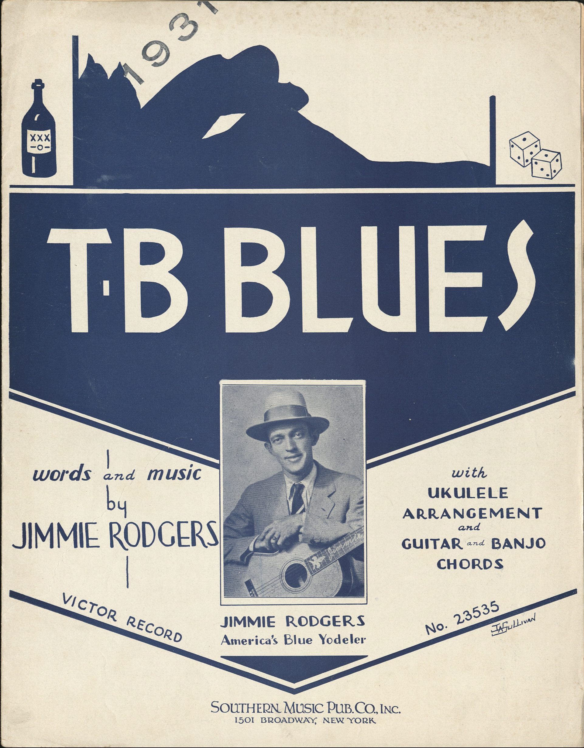
The above lyrics come from the 1931 song, “T.B. Blues” by pioneering country singer, Jimmie Rodgers. They describe a young man accepting his fate and losing the fight against tuberculosis, or TB.
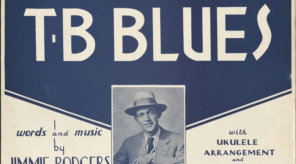
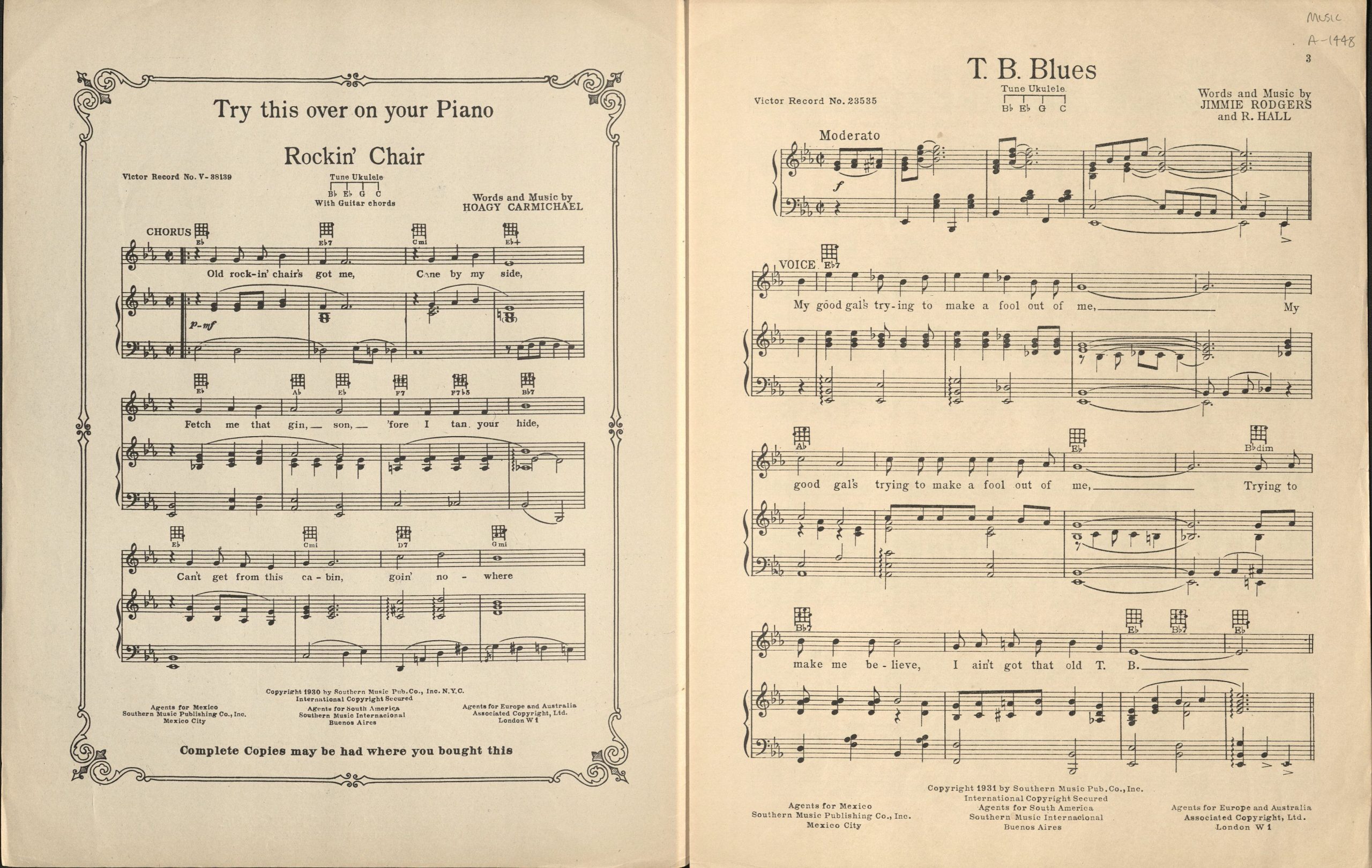
By the 20th century, sheet music had long been a tradition in the music industry as a way for customers to immerse themselves with their favorite songs but also an opportunity for companies to advertise their artists and products. The sheet music for “T.B. Blues’” was published in 1931 by the Southern Music Company – though it was based in New York City. It includes the then standard “Try it on your Piano” introduction page and advertisements for other Jimmie Rodgers songs and that of the Carter Family and band-leader Hoagy Carmichael. The front cover features a portrait of Jimmie Rodgers in his signature suit and straw hat, under a banner with the song title and the curious inclusion of a moonshine bottle, a pair of dice, and the silhouette of a man laying in bed with his chest to his knees, perhaps an allegory to the pain suffered by tuberculosis patients.
Known as the “Singing Brakeman,” a reference to his time working on railroad lines, Rodgers is considered the “father of country music” for his influence across country, rhythm and blues, bluegrass and rock n’ roll. An inductee of the Rock n’ Roll Hall of Fame and Country Music Hall of Fame, Rodgers is known for such hits as “In the Jailhouse Now,” “Blue Yodel No. 9” (with Louis Armstrong) and “T for Texas,” and has been covered by legendary artists Bob Dylan, Willie Nelson, Johnny Cash and Allison Krauss, among others. Tragically, his promising career lasted only six years and was cut short after a long battle with tuberculosis, when he succumbed to the disease in 1933, at the age of 35.
Sheet Music for T.B. Blues (right), including the “Try this over on your piano” intro page.
Got me worried soul, I can’t even sleep at night
I’ve got the T.B. blues
“TB” BACKGROUND
In Jimmie Rodgers’ lifetime, tuberculosis was “one of the two leading causes of death in the early 1900s” and the “dominant chronic infectious disease of the first half of the twentieth century.” Tuberculosis — known also as consumption, phthisis, white plague, and “the robber of youth” throughout history — is caused by the bacteria, mycobacterium tuberculosis.
The earliest written description of TB dates back three millennia to ancient India and in AD 174, the Greek physician Galen described its symptoms as “fever, sweating, coughing and blood stained sputum.” Thought to be hereditary until the late 19th century, German scientist, Robert Koch, discovered that tuberculosis was an airborne infectious bacterial disease that could be transmitted from person to person.
According to a study by Harvard University Library, tuberculosis caused more deaths in industrialized countries than any other disease during the 19th and early 20th centuries. However, a romanticized view of tuberculosis had sprung up in the 1800s as the disease came to be associated with artists and literature. Some believed that suffering from the disease increased creativity, “heightened sensitivity and spiritual purity.” Writer Robert Louis Stevenson suffered from tuberculosis for most of his life and artists such as Emily Bronte, John Keats, and Frederic Chopin all died from the disease at an early age.
Alexander Dumas claimed, “It was the fashion to suffer from the lungs; everybody was consumptive, poets especially; it was good form to spit blood after each emotion that was at all sensational” while Lord Byron quipped that he “should like to die of consumption”.
Yet for many suffering from tuberculosis, the disease could feel like a slow death. Tuberculosis can attack the body in different ways, from the lungs to the kidneys, brain and spine. TB bacteria can settle in the lungs and begin to grow and move through the blood to other parts of the body. Not all who contract tuberculosis become sick leading to the distinction between Latent TB Infection (asymptomatic) and TB Disease (symptomatic).
Like many who suffered from the progressive form of tuberculosis, Rodgers’ battle was prolonged and extremely painful, coughing up bloody sputum for years and suffering from chronic fatigue. At the time of the recording of “T.B. Blues” in 1931, Rodgers had already been living with the disease (the symptomatic TB Disease) for over seven years. He had been diagnosed in 1924 by a family physician after suffering a hemorrhage (Porterfield, p. 53).
When it rained down sorrow, It rained all over me
‘Cause my body rattles, Like a train on that old S.P. [Southern Pacific RR]
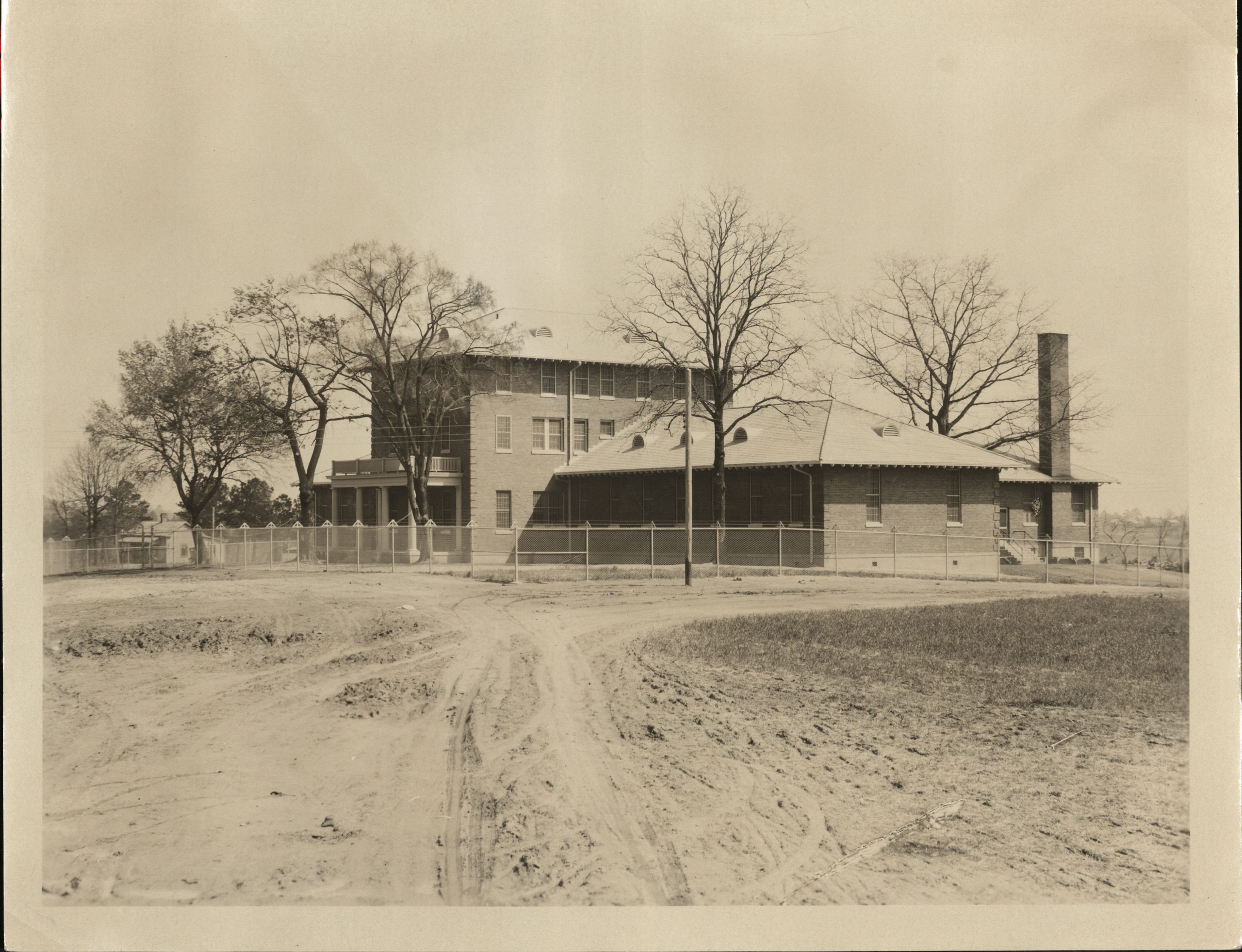
Image of the Durham County Jail, circa 1920s. Building and grounds would be converted to TB Sanatorium in 1943 and 1944.
Prior to the innovations of vaccines, medication, and antibiotics that have helped fight tuberculosis, most physicians could only prescribe a nutritious diet, rest and fresh air. In the late 1800s and early 20th century, tuberculosis sanatoriums were established throughout the United States and Europe where TB patients could isolate and rest.
However, relaxation, bedrest, quarantine and sanatorium care weren’t necessarily options for those suffering from poverty. Not working meant not getting paid, and the same was true for Jimmie Rodgers. Particularly for a musician just reaching stardom, taking time from work was not an option. He spent time in sanatoriums and even lived in Asheville, NC for its cooler climate and mountain air; but he continued to perform, even against the recommendations of physicians and family (Porterfield, p.53). Rodgers would on occasion stumble out of bed to perform while fighting a fever and went so far as to tape plaster to his ribs to dull the pain and prevent from breathing too deeply. When he couldn’t stop coughing onstage, fans were known to applaud sympathetically and shout, “Spit ‘er up, Jimmie and sing some more” (Porterfield; p. 115; 279).
Eventually, the disease and its complications would prove too much. Jimmy Rodgers lost his battle with tuberculosis on May 26, 1933. Ever the tireless performer, Rodgers spent his final days recording music in a New York studio, cutting his last record two days before his death.


 The 1928 “little library” by White is a series of 28 books whose length ranges from 20–48 pages. While small, I would say that calling them “thumb-nail” editions is a little misleading; the books measure at 4.5 inches in height and near 3.5 inches across (3 ⁷⁄₁₆ to be exact) is far from what is considered a miniature book or thumbnail sized. The advertisement at the back for each book boasted that each book contained illustrations, sometimes in color, and provided White’s sound advice on “health building by natural living.” Each book could be purchased for 25 cents (now somewhere near $4.50) or, for 5 dollars prepaid (around $90 for us today), one could score for the entire set.