

Submitted on behalf of Erin Hammeke
For conservators, one of the aspects of having the MSI system that excites us most is being able to visualize and document the effects of the treatments that we perform. Although we are still learning the ropes with our new system, we had a recent opportunity to image some iron gall ink documents. Iron gall ink is common historic ink that reacts with moisture in the environment to form acidic complexes that spread and sink into the paper, weakening the paper and, in some cases, leaving holes and losses. This iron gall ink degradation can be better visualized with MSI, since the beginning stage, haloing, is not always visible under normal illumination. Look here for more information on iron gall ink damage and here for using MSI to document iron gall ink condition and treatment. We also illustrated the haloing effect of iron gall ink damage using MSI on Jantz MS #124 in a previous post.
Recently, DUL conservators experimented with treating some discarded iron gall ink manuscripts with a chemical treatment that aims to arrest the ink’s degradation. This treatment requires submerging the manuscripts in a calcium phytate solution – a chemical that bonds with free iron (II) ions, stabilizing the ink and preventing it from corroding further. The document is then rinsed in a water bath and an alkaline reserve is applied. Resizing with gelatin is another common step, but we did resize our test manuscripts.
Since these were discarded test material, we were able to cut the manuscripts in two and only treat one half. Imaging the manuscript with MSI revealed some notable findings.
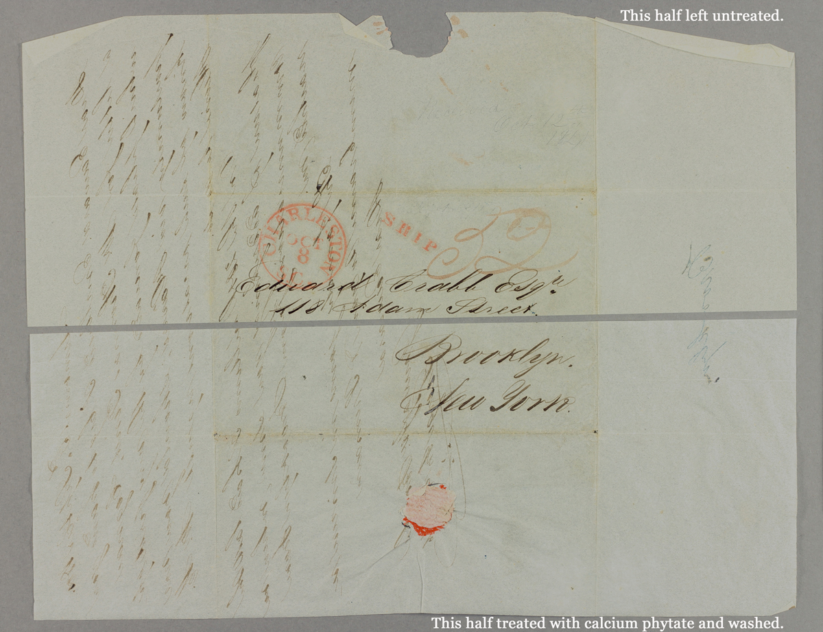

Most of the treated papers now appear lighter and brighter under normal illumination because they have been washed. However, the untreated halves exhibited pronounced UV induced visible fluorescence around the 488 nm range and the treated halves did not. We believe this difference likely has to do with washing the paper substrate and rinsing out degradation products or perhaps paper size that may exhibit fluorescence at this wavelength. We were happy to see that for a treatment that targets the ink, there was very little noticeable difference in the appearance of the inks between untreated and treated portions of the test manuscript. There was some reduction in the “ink sink” (ink visible from the opposite side of the manuscript) and a very slight softness to the edges of the ink in the treated sample, but these changes were very minimal. We look forward to imaging more of our test manuscripts in the future and seeing what else we can learn from them.
______
Want to learn even more about MSI at DUL?
- Watch an imaging Session
- Read other MSI posts on Duke Libraries’ Bitstreams and Preservation Underground blogs