descriptive image

Enticing Engineers: New Areas of Outreach in the Rubenstein Library

Post contributed by Rachel Ingold, Curator for the History of Medicine Collections.

In September, the Rubenstein Library partnered with colleagues in the Natural and Engineering Sciences (NSE) for an open house event. While our Engineering Exposition targeted students, faculty, and staff from Duke’s Pratt School of Engineering, all were welcome to attend.

Photograph showing a large room with several tables displaying books. At one table, three people look closely at two books.
Faculty from the Engineering School examine works on engineering from the 16th and 17th centuries! Photo by Deric Hardy.
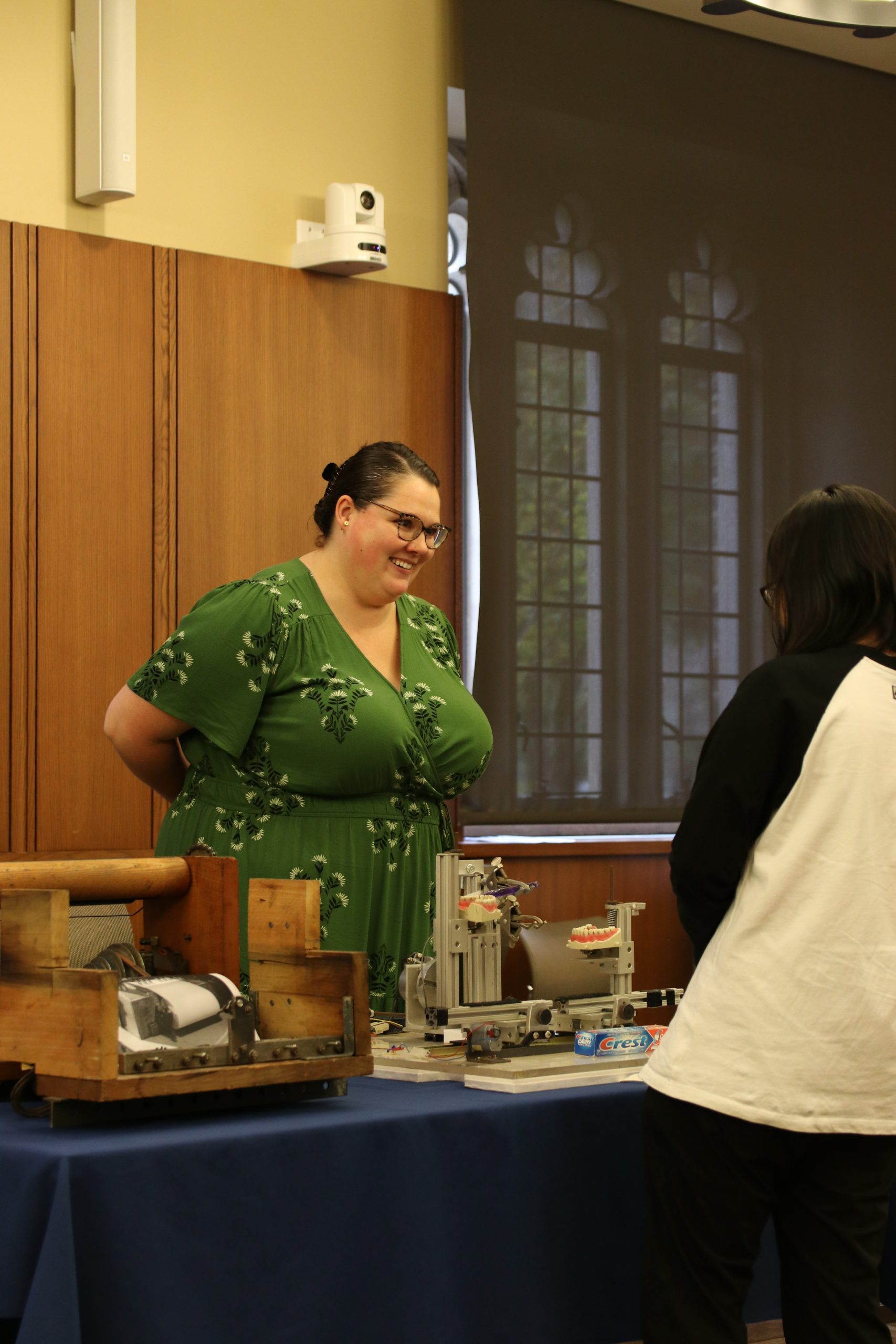
A woman with brown hair wearing a green dress talk with a student, whose back is toward the camera. On a table between the two people, is a toothpaste testing device composed of metal parts and sets of fake teeth.
Robin Klaus, graduate Intern in the Hartman Center for Sales, Advertising, and Marketing History talks with a student about the toothpaste testing device found in the Consumer Reports archive. Photo by Janelle Hutchinson.


Items from a variety of collecting areas within the Rubenstein Library were available for visitors to examine and handle. Some highlights included

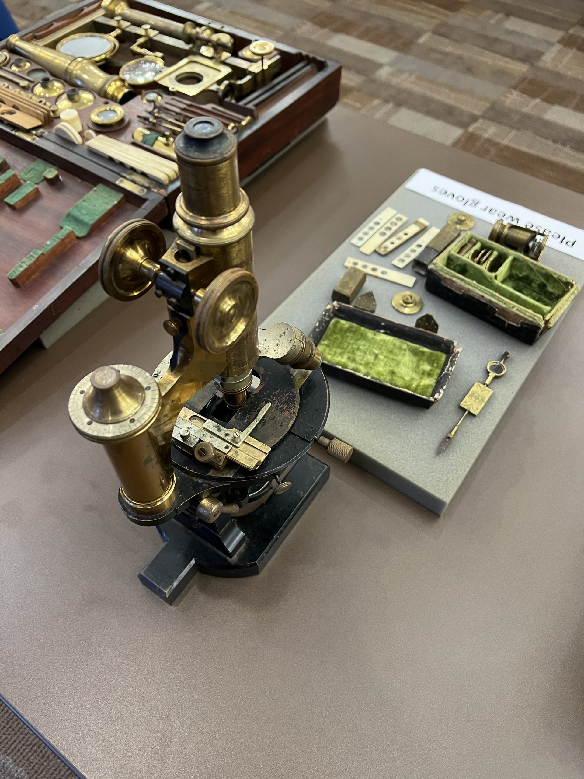
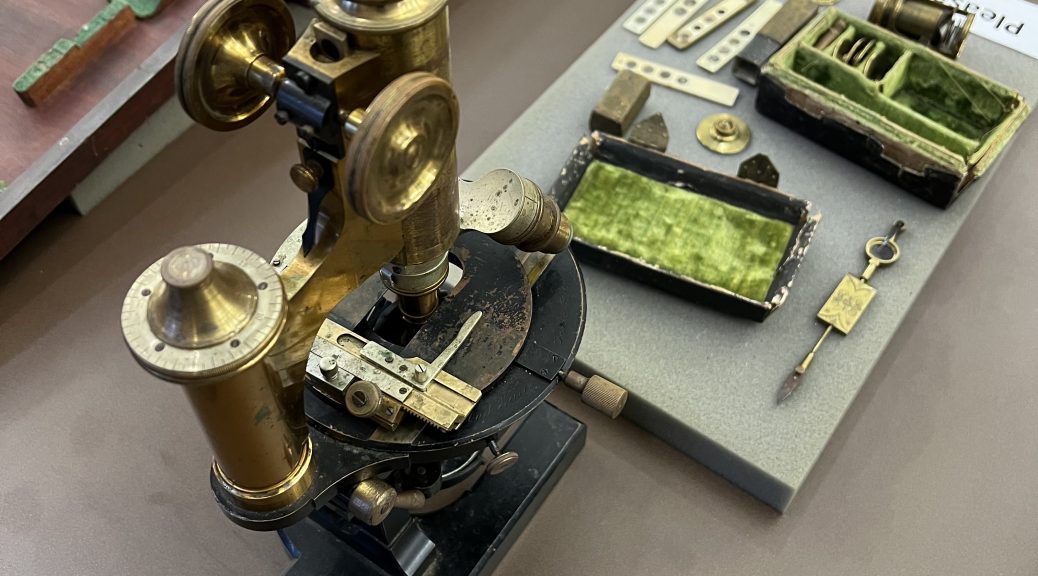
Photograph of a table with three examples of microscopes on display.
Examples of 18th and 19th century microscopes that visitors were encouraged to handle and try out! Photo by Deric Hardy.
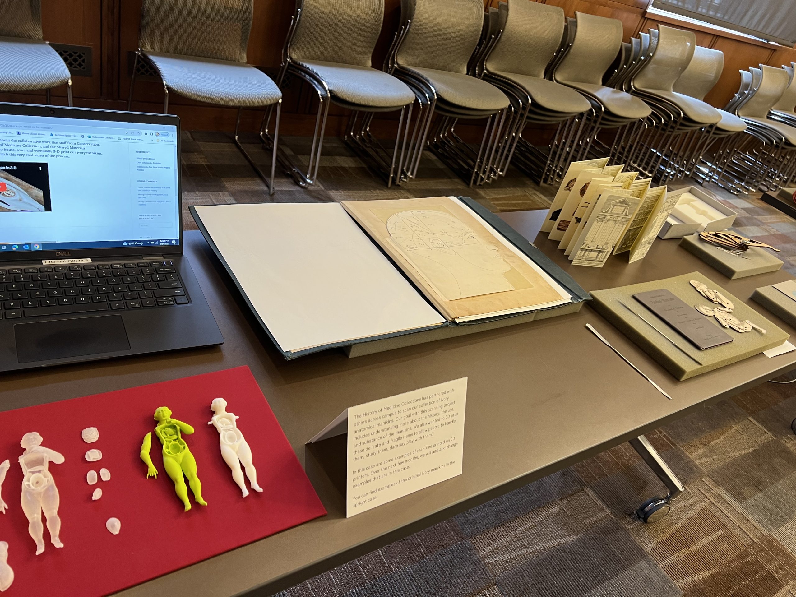
Photograph of table displaying several small, plastic anatomical figures as well as a laptop playing a video showing the making of those figures. The table also displays several books featuring pop-up components.
Examples of moveable books from the History of Medicine Collections and samples of 3D printed anatomical manikins made from items in our collection! Photo by Deric Hardy.
Image from an anatomical flap book. The left side of the image shows an artistic representation of a human eye and human ear and shows the flaps closed. The right side of the image shows the paper flaps lifted to reveal, underneath the original images, the anatomy of the inside of the eye and inside of the ear.
Pages from an anatomical flap book where the flaps can be lifted, as shown on the right, to reveal detail about the human body. Photo by Janelle Hutchinson.

Anyone is welcome to view our items in-person during our open hours. We also have great digital collections.

We look forward to our continued partnerships with colleagues across the Library and campus. Let us know what you might like to see at our next Engineering Exposition!