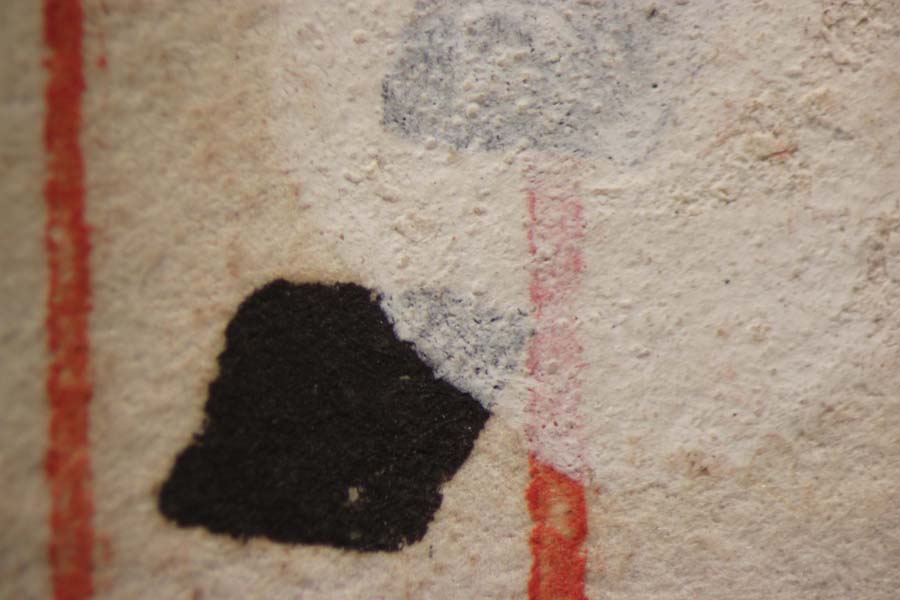
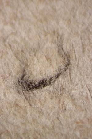
We got a new toy camera for our microscope this month. The last one didn’t really work well, something to do with the adapter ring and our Nikon. This one, a Canon, is designed to work with our particular scope and came with the correct adapter and software. We can now easily make documentation images of friable media, mold spores…you name it. Here are some sample test shots I took ten minutes after setting it up.


We still need to learn how to tweak the depth of field and optimize the exposure set points. But already this camera is far superior to our old set up. Having the ability to take close-up images is important in order to identify areas of damage and to document before- and after-treatment conditions. We look forward to using it as an integral part of our treatment protocol.

