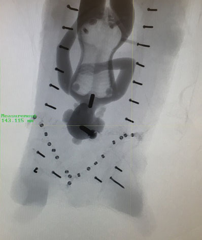
Conservator Erin Hammeke has been working with History of Medicine Curator, Rachel Ingold and SMIF Research and Development Engineer, Justin Gladman to facilitate the scanning of our 22 ivory manikins using a High Resolution X-ray Computed Tomography Scanner (Micro CT scanner). These high resolution 3-D scans allow us to see internal components of the manikins, thoroughly document them and their component parts, and also to create 3D printed models to allow for unfettered access and handling of surrogate manikins by patrons. So far, we have imaged over half of the collection of 22 manikins to date over 7 imaging sessions.
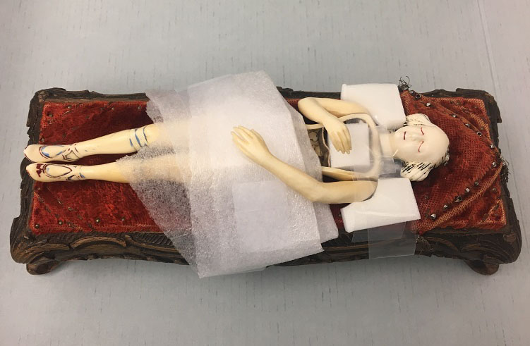
Conservation’s support for this project has been a team effort. First we researched the safety of the process for ivory and component parts. Then we thought through the logistics of ensuring their physical safety and security during imaging. Over the past year, Conservation Specialist, Rachel Penniman has been carefully boxing each of the manikins in terrific custom padded artifact boxes (see Quick Pic: Boxing Near-Naked Ladies) to assist with their safe transport to the SMIF facility on campus. After transport to SMIF, Erin unpacks the manikins and removes their parts before securely wrapping them in low density material to support the manikin during the 20-30 minute scan. Thanks to Beth for sewing custom foam supports for this project!
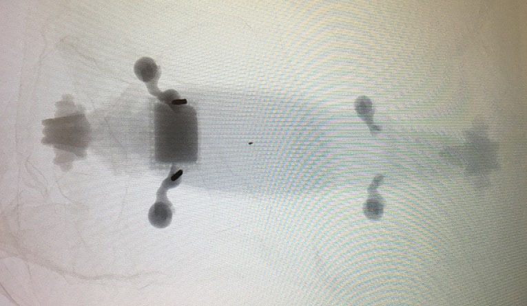
Smaller, removable internal organs are imaged in separate scans to enable an adjustment to the scanning resolution and isolation of the component parts in the digital scan and 3D print.

This photos shows some of the ways in which Erin strapped and supported a manikin with a loose arm during scanning, as well as still images from the scan that show internal metal fasteners and repairs that are not visible upon external examination.
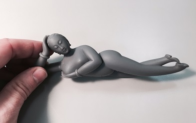
And here is an example of a 3D print of one of our solid manikins showing fantastic detail.
Keep an eye out for a more in depth Duke News story about the project by science writer Robin Smith, PhD.


