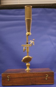

With generous assistance from the History of Medicine travel grant, I traveled to Duke University to view and photograph historical obstetric and gynecological tools housed in Duke’s History of Medicine Collections at the Rubenstein Rare Book & Manuscript Library. There I viewed various artifact collections donated by practicing regional doctors, including the L. M. Draper Collection, the George D. & Evelyn Wilbanks Collection, and several anonymous collections. I also viewed anatomical lift-the-flap guide books, lift-the-flap anatomical fugitive sheets and the Trent Collection of Ivory Anatomical Manikins, all of which were used to teach medical procedures, including delivery.
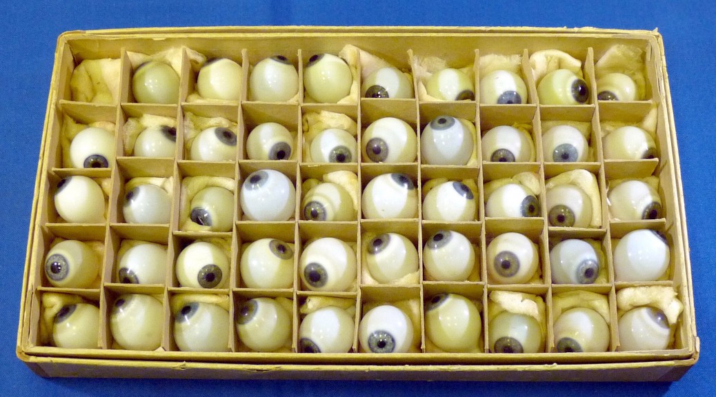
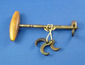
Having access to Duke’s collection was an incredible experience. I treated it like a short artist residency. I set up my lights, a pop-up tent, my camera and a tripod in a study room within the library. Every morning, a cart was wheeled in with OB/GYN tools, anatomy text books and glass slides. It was exciting (and a little nerve-wracking), opening up boxes and not knowing their contents. For some items, I felt I was discovering the files for the first time. In a way I was: besides the archivists who received and catalogued them, some of the items had never been requested. I often felt as though I were in the medical field—donning nitrile gloves, carefully removing the items from their boxes, gently lying them down on the fabric of my pop-up lighting tent, careful not to harm them in any way. I found myself photographing them as abstractions or as jewelry, a style of cataloguing unlike other projects I have photographed.

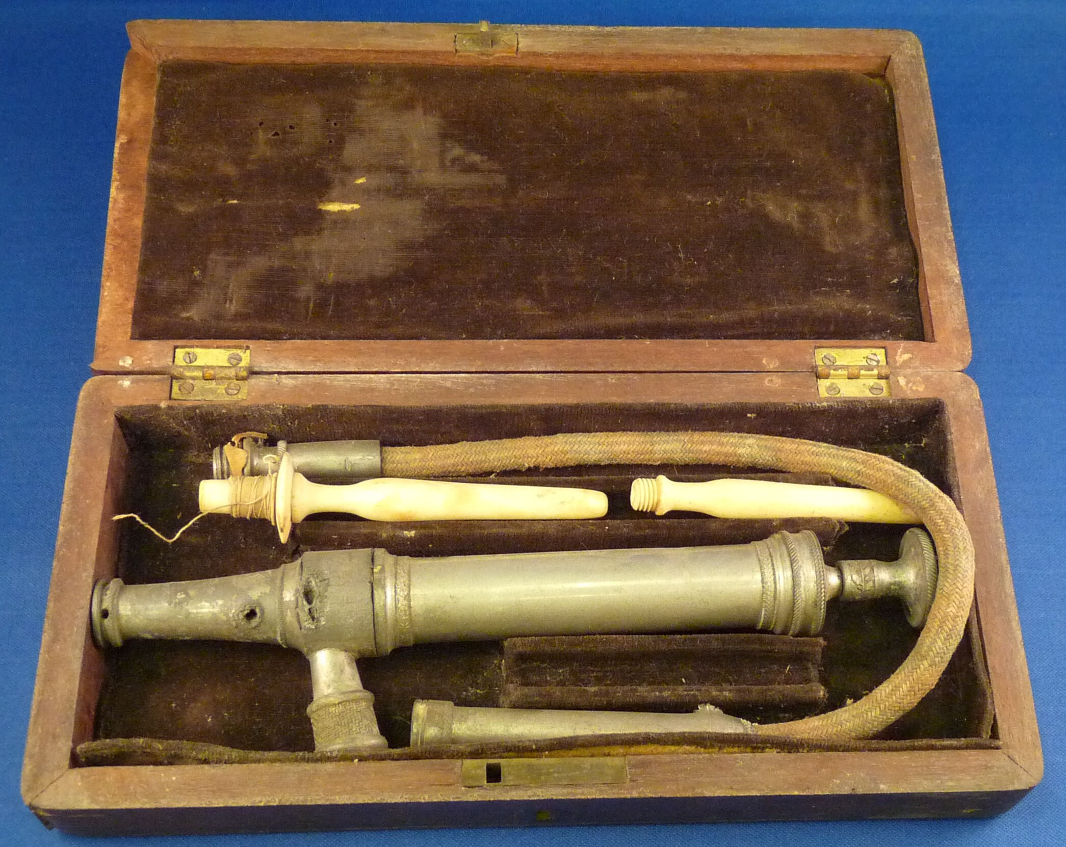
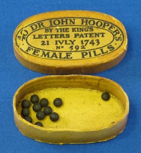
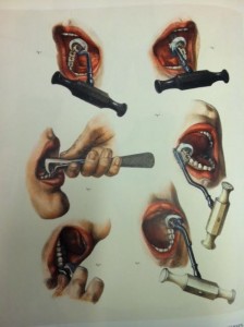
My work focuses on historical and contemporary women’s lives and I am particularly interested in the past’s technology and how it relates to today. I have previously done photographic projects on antique vibrators, social media and the practice of keeping a commonplace book and with this project, the history of labor and delivery technology. While the process of getting pregnant has changed with IVF and the location of delivery may have changed, the actual process of delivery has not changed. Although American society emphasizes new products & experiences, and the medical world uses recent technology & procedures, women continue to deliver only one of two ways—vaginally or via Cesarean section. Prior to my arrival at Duke, I assumed the tools used in labor and delivery were harmful to the infants and delivering women. I also wondered how deadly labor actually was—in fictionalized accounts in both books and screen, no female who delivered a newborn ever lived, and seldom the child. I expected antique tools to be brutal and different in appearance than today. It surprised me that many of the tools I photographed resembled contemporary tools, only with time’s effect through rust or evident aging.
My research at Duke is the beginning of both my project and into further research on the history of the OB/GYN tools and their uses. Although in its early stages, I plan to study these tools’ history, as well as their use & influence today. The final images may be printed as slides, emulating turn of the twentieth century magic-lantern plates or late-twentieth century educational slide shows. Whatever form these images take, I was particularly inspired by the anatomical lift-the flap books & broad sides and will create an artist book influenced by these interactive educational guides. I look forward to sharing future developments of this project. Thank you to everyone at Duke University’s Rubenstein Library for their assistance during my stay.
Post contributed by History of Medicine Travel Grant recipient Lindsey Beal. Beal is a photo-based artist and professor in Providence, Rhode Island. Her work and further information can be found at lindseybeal.com.