Tag Archives: anatomy

A Melancholy Skeleton

Post contributed by Roseen Giles, PhD, Assistant Professor of Music and Curator, Duke University Musical Instrument Collections

A white ivory carving against a black velvet background. The carving shows a skeleton leaning against a pedestal with a clock. Symbols of worldly power and wealth--like a crown, a knight's helmet, and flags--are piled at his feet.
Memento Mori, approximately 1650, Rubenstein Library

There is a melancholy skeleton who lives in Duke University’s History of Medicine Collections at the Rubenstein Library. In truly every sense, he is a marvel. Meticulously carved from a single piece of ivory, this piece is crafted with astounding virtuosity by an artist who must have known as much about the science of anatomy as they did of the human condition. The composition dates from the middle of the seventeenth century but the name of the artist and the circumstances of its creation are unknown. It is perhaps fitting that the subject of this skeletal wonder is the greatest mystery of all: death. The remarkable details of the carving suggest that it is a vanitas or memento mori, a genre of artworks that remind us through a series of recognizable symbols of the certainty of death and the fleetingness of life. The skeleton itself represents a human life spent; despite our differences and all the things that mark us as unique individuals we will all one day die, leaving behind ossified remains that are, to the untrained eye, indistinguishable one of from the other.

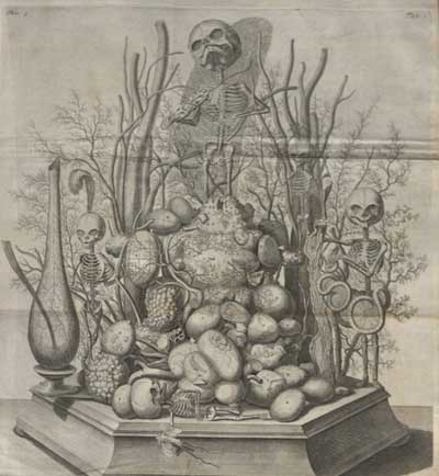
At the skeleton’s feet are some of the most common symbols of the vanitas genre: traces of earthly pursuits such as warfare, monarchy and, perhaps most importantly, objects which point to the passing of time. This level of detail will be familiar to those acquainted with Hans Holbein the Younger’s masterpiece The Ambassadors (1533), a painting which contains quintessential examples of vanitas, among them a lute with a single broken string. Music and sound can in this case stand in as a metaphor for human life, one that is equally transient and decaying as soon as it is brought into existence. Tucked behind our skeleton’s crossed legs we can see a sickle or scythe, the symbol for the tyranny of time and of its god Saturn. The pedestal upon which he rests his arm contains the mechanical parts of a timepiece, innovative new inventions in the early modern period. The skeleton cradles his own head as he leans upon the ticking pedestal in a striking gesture that suggests both a contemplative and melancholy temperament. Perhaps most astonishing is the detail of the sash that surrounds but does not fully touch his back—as if it had been taken up with a momentary gust of wind that froze it in its movement.

Black-and-white illustration of a skeleton leaning on a shovel. Its skull is tilted back and its mouth is open. There is a mountainous landscape behind it.
Andreas Vesalius, De humani corporis fabrica (1543), p. 163

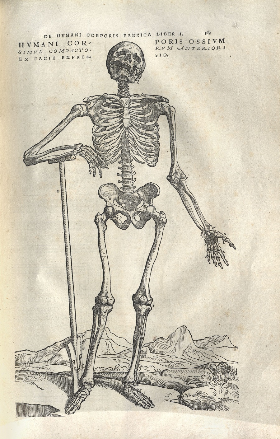
The stick upon which the skeleton rests his right hand does not seem particularly remarkable, but a comparison to two drawings from Andreas Vesalius’s (1514–64) revolutionary anatomical treatise De humani corporis fabrica [On the Fabric of the Human Body] (Basel, 1543) reveals that there may be more to this detail than meets the eye. In one of these illustrations—possibly attributable to a pupil of Titian, Joannes Stephanus of Calcar (c. 1488–1576)— a skeleton turns his head up towards the heavens (Figure 2). The positioning of his neck and torso suggests a depth of feeling: his head is turned slightly to one side as he looks up and he rests his weight on a staff very much like the one in the ivory vanitas. In Vesalius we can see that the skeleton is leaning not on a walking stick but on a shovel, which he will use to dig his own grave. In yet another of the images from the Fabrica we can see the same skeleton looking not upwards but down with one hand supporting his own head and the other resting on a skull—not his own, apparently— which is placed upon a pedestal very similar to the timepiece in our carving. The anatomical accuracy in both the drawings of the Fabrica and of our ivory skeleton is remarkable enough, but their gestural embodiment of human thought and emotion make these works truly astonishing historical witnesses. Their subjects point to the contradictions of lived existence: the unstable dichotomy between the physical and mental, between anatomy and emotion, between scientific and artistic knowledge.

Black-and-white illustration of a skeleton leaning on a tomb. It rests its skull on its left hand and places its right hand on another skull.
Vesalius, De humani corporis fabrica (1543), p. 164

The unknown artist who made the vanitas skeleton in Duke’s History of Medicine Collections understood the rhetorical power of the ephemeral made material, when the truths of existence are brought into dialogue with the irrationality of consciousness. The very material they chose is telling of this. Ivory is rare and costly but lends itself well to the intricate detail of the piece’s low relief; it is not human bone but stands in for it in a vivid and arresting way. There are many things that such a piece can teach us about the history of medicine and, more specifically, seventeenth-century understandings of human anatomy and the physicality of emotions. But in reminding us that we will certainly die it can also tell us something about how to live. It represents, in short, a kind of contemplative interaction between the living and the dead, an interaction which suggests that the health of the human body is inextricably linked with our thoughts and emotions. In this way our melancholy skeleton can, even by representing death and transience, help us to understand and to improve the health and well-being of the living.

Frederik Ruysch’s Anatomical Art

Post contributed by Amelia Holmes, Josiah Charles Trent Intern for the History of Medicine Collections.

Anatomical specimens emerged as an art form near the end of the seventeenth century. Although they may seem morbid today, at the time of creation, they were viewed as striking a balance between the scientific and the artistic. They served to educate people on human anatomy as well as to remind them of the fleeting nature of life.

Illustration from Opera Omnia Anatomico-Medico-Chirurgica, ca. 1737.

One of the more notable creators of anatomical art is Frederik Ruysch, a Dutch botanist and anatomist who lived from 1638 to 1731—an impressive 93 years in a time when many died young. A capable researcher, Ruysch was the first to describe bronchial blood vessels, the vascular plexus of the heart, and the valves of the lymphatics. However, his real interest lay in anatomical preparations, and he has been described by a recent biographer as “probably the most skilled and knowledgeable preparator in the history of anatomy” (Gould, p. 20). Ruysch served as the chief instructor to midwives and the “legal doctor” to the court of Amsterdam. Through these positions, he had easy (and legal) access to the bodies of stillborns and dead babies.

The preparations were initially created to use in his classes, but they eventually gained an interest from the public. To showcase his vast collection (he created more than 2,000 from 1665 to 1717 alone), he opened his own cabinet of curiosities to the public, which for many marked the first time they were able to see human internal organs. The collection was also noteworthy because of the lengths to which Ruysch went in an effort to make the specimens appear more natural. For example, embalmed children were clothed or held bouquets of preserved flowers. In 1717, Peter the Great, who was an admirer of Ruysch, purchased the entire cabinet of curiosities for 30,000 guilders. The collection was then shipped to St. Petersburg, and along with the cabinet of curiosities formed by Albertus Seba, they became the core of the Kunstkammer—the Academy of Sciences of Russia’s first public museum.

Although a number of Ruysch’s wet preparations still exist today (a fact which he would find unsurprising), none of his dry specimens have been located. He used fetal skeletons and other body parts to create multi-specimen scenes. These scenes served as the centerpieces for each of the literal cabinets within the rooms of his museum. As Gould points out, these tableaux were focused on allegorical themes such as death and the transience of life. The small skeletons are decorated with symbols of death and short life: mayflies rest in hands, skulls weep into handkerchiefs made of mesentery, and snakes made of intestine wine their way through bones. Today, these still-life scenes exist for us only through second-hand descriptions and, fortunately, through a number of engravings.

Fortunately for those interested in seeing these illustrations up close, the History of Medicine Collections has two volumes from the multi-volume Opera omnia anatomico-medico-chirurgica. On October 29, from 2-4 pm, they will be on display as part of Screamfest in the Holsti-Anderson Family Assembly Room.

Recommended Reading:

New Acquisitions: Unique Depictions of the Human Body

In June and July we’re celebrating the beginning of a new fiscal year by highlighting new acquisitions from the past year. All of these amazing resources will be available for today’s scholars, and for future generations of researchers in the Rubenstein Library! Today’s post features a new collection in the Library’s History of Medicine Collections. Check out additional posts in the series here.

fugitive sheet female

The History of Medicine Collections has acquired two anatomical fugitive sheets, elevating our holdings to now include ten of these magnificent items. Anatomical fugitive sheets are single sheets, similar to broadsides, that are printed on one side. Illustrations of the human body accompany text that was written in Latin, and later in the vernacular. Dating from the sixteenth century, this pair of fugitive sheets, titled Viscerum hoc Est Interiorum Corporis Humani Partium Descriptio and published in Antwerp in the sixteenth century, includes hand colored illustrations with accompanying text in Latin.

Besides being incredibly rare—these are the only known copies of these sheets—the sheets are noteworthy for many reasons, including the depiction of the human body using three-dimensional flaps that lift to reveal internal organs, as the title suggests.  This particular pair of fugitive sheets has lost most of its flaps. While the male figure only retains a fragment of one flap, the female figure retains one full flap of the inner organs in entirety. Such loss is common since most of these fugitive sheets date to the sixteenth and seventeenth centuries and were printed as single sheets.

Theories abound as to who would own such items. Were they created for physicians, barber surgeons, or the lay person wanting to know more about the human body? Were they hung in apothecaries, medical university classrooms, or the gentleman’s library? Any sheets that remain today are incredibly rare and worthy of study and analysis. These appeal not only to the medical student who wants to see what inaccuracies exist, but to those interested in the history of science, printing history, and art history.

Post contributed by Rachel Ingold, Curator, History of Medicine Collections.

New Acquisitions Week, Day One: Moveable Brains and Laughing Cows

We’re celebrating the beginning of a new fiscal year with a week’s worth of new acquisitions from the first half of 2012.  Two newly acquired selections will be featured in a post every day this week.  All of these amazing resources will be available for today’s scholars, and for future generations of researchers in the Rubenstein Library!

Diagram of the brain, from Ludwig Fick, Phantom des Menschenhirns (1885).
  • Joy Golden Papers: Joy Golden was a well-known advertising copywriter who started her own creative company, Joy Radio, in the 1980s that specialized in humorous radio advertising. She did a series of commercials for Laughing Cow Cheese that became particularly well known.  She also was active in the Friars Club, including holding the position of Governor.  Her papers include files related to her work in advertising from the 1960s forward, and audiotapes of many of the radio advertisements created by her company.  Her papers add to the Hartman Center for Sales, Advertising, and Marketing History‘s rich collections on women and advertising and the development of radio advertising.